Carbon Accumulation in Lung Cells Linked to Severity and Inflammation in Chronic Obstructive Pulmonary Disease Patients

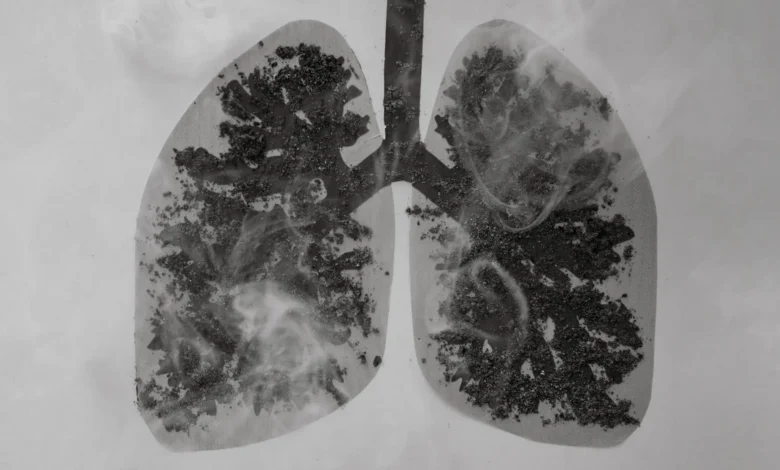
A landmark study published in the journal ERJ Open Research has revealed a significant disparity in the accumulation of soot-like carbon deposits within the lung cells of individuals suffering from chronic obstructive pulmonary disease (COPD) compared to those who smoke but have not developed the condition. The findings, released on June 10, provide a critical new perspective on how environmental pollutants, ranging from cigarette smoke to diesel exhaust and industrial emissions, interact with the body’s internal defense mechanisms to exacerbate respiratory decline.
The research, conducted by a team of scientists at the University of Manchester, suggests that the inability of specific immune cells to clear carbon may be a defining characteristic of COPD progression. By examining the cellular architecture and waste-clearing efficiency of alveolar macrophages—the "scavenger" cells of the lungs—the study highlights a direct correlation between carbon buildup, cellular inflammation, and the loss of lung function.
Understanding the Role of Alveolar Macrophages
To comprehend the significance of this study, it is necessary to understand the primary function of alveolar macrophages. These specialized white blood cells reside on the internal surfaces of the air sacs (alveoli) in the lungs. Their primary responsibility is to identify, engulf, and neutralize foreign particles that bypass the upper respiratory tract’s filtration systems. This includes bacteria, viruses, and particulate matter such as carbon.
Under normal circumstances, these cells act as the first line of defense, preventing pollutants from causing systemic damage or localized infection. However, the Manchester study indicates that in patients with COPD, this defense mechanism appears to be compromised. Instead of clearing the carbon, the macrophages become repositories for it. As carbon accumulates, these cells undergo physical and functional transformations, growing larger and paradoxically triggering the very inflammation they are meant to prevent.
Study Methodology and Comparative Analysis
The research team, led by Dr. James Baker and Dr. Simon Lea, utilized a rigorous comparative framework to isolate the effects of carbon accumulation. The researchers obtained lung tissue samples from 43 individuals undergoing surgery for suspected lung cancer. To ensure the integrity of the data, the researchers only analyzed portions of the tissue that were free of malignant cells.
The cohort was divided into two distinct groups: 28 patients diagnosed with COPD and 15 individuals who were current or former smokers but did not exhibit the clinical markers of COPD. This distinction was vital for determining whether carbon accumulation was merely a byproduct of smoking or a specific pathological feature of COPD.
Using advanced microscopic techniques, the team measured the physical dimensions of the alveolar macrophages and quantified the volume of carbon deposits within them. The results were stark: the average amount of carbon found in the macrophages of COPD patients was more than three times higher than the levels found in the smokers without the disease. Furthermore, the study observed a consistent trend where cells laden with carbon were significantly larger than those without visible deposits, suggesting a physical strain on the cellular structure.
The Correlation Between Carbon and Lung Function
One of the most critical aspects of the study was the link between cellular carbon levels and clinical respiratory health. The researchers utilized a standard diagnostic metric known as FEV1% (Forced Expiratory Volume in one second). This test measures the volume of air an individual can forcefully exhale in a single second, serving as a primary indicator of airway obstruction and overall lung capacity.
The data revealed an inverse relationship: patients with the highest concentrations of carbon in their alveolar macrophages recorded the lowest FEV1% scores. This suggests that the accumulation of carbon is not just an incidental finding but is closely tied to the severity of the disease. When the researchers replicated these conditions in a laboratory setting by exposing healthy macrophages to carbon particles, the cells responded by expanding in size and secreting high levels of pro-inflammatory proteins. This laboratory phase confirmed that the presence of carbon directly stimulates a chronic inflammatory state within the lung tissue.
Expert Perspectives on the "Clearing" Hypothesis
The study’s findings raise a fundamental question regarding the etiology of COPD: why do these patients accumulate more carbon than smokers without the disease? Dr. Simon Lea, one of the study’s lead authors, posited two primary theories. The first suggests a physiological "clearance" failure. In this scenario, individuals who develop COPD may have a genetic or acquired inability to effectively transport carbon-laden macrophages out of the lungs.
The second theory focuses on environmental exposure thresholds. It suggests that individuals exposed to higher concentrations of particulate matter over a lifetime may reach a "tipping point" where the macrophages become overwhelmed, leading to the onset of COPD. "We can see that this build-up of carbon is not a direct result of cigarette smoking alone," Dr. Lea noted. "Instead, the macrophages in COPD patients are inherently different in their form and function. They are less capable of maintaining a clean environment within the alveoli."
Dr. James Baker emphasized that while COPD is a complex disease influenced by various genetic and environmental factors, the role of air quality cannot be overstated. "We wanted to study what happens when this carbon builds up, as it may influence the cells’ ability to protect the lungs," Baker stated. The study confirms that this buildup is a hallmark of the diseased lung, potentially serving as a driver for the chronic inflammation that defines the condition.
Broader Implications for Public Health and Policy
The implications of the Manchester study extend far beyond the laboratory, touching on urban planning, industrial regulation, and public health initiatives. Professor Fabio Ricciardolo, Chair of the European Respiratory Society’s group on monitoring airway disease, who was not involved in the study, noted that the research provides a missing link in our understanding of how polluted air worsens respiratory conditions.
"This build-up seems to be altering those cells, potentially causing inflammation in the lungs and leading to worse lung function," Ricciardolo remarked. He further argued that the findings provide a scientific basis for more aggressive air quality standards. As diesel exhaust and industrial soot are major sources of the carbon found in these cells, reducing particulate matter in urban environments could potentially slow the progression of COPD in vulnerable populations.
The study also reinforces the necessity of smoking cessation programs. While the research showed that COPD patients have higher carbon levels than non-COPD smokers, smoking remains a primary delivery system for carbon particles. By quitting, patients can at least stop the influx of new deposits, though the study suggests that the carbon already present may remain "trapped" in the macrophages for extended periods.
Future Research and Clinical Outlook
The discovery that carbon-laden macrophages promote inflammation opens new avenues for therapeutic intervention. Current COPD treatments largely focus on bronchodilation—opening the airways—and reducing symptoms. However, this research suggests that targeting the macrophages themselves, or the inflammatory proteins they produce in response to carbon, could lead to treatments that address the underlying cellular dysfunction.
Future studies are expected to focus on the long-term behavior of these carbon deposits. Scientists want to determine if there is a way to "re-activate" the clearing mechanism of the macrophages or if specialized medications can neutralize the inflammatory signals sent by carbon-clogged cells. There is also a growing interest in using carbon accumulation as a biomarker; by measuring the soot levels in a patient’s lung cells, doctors might one day be able to predict the rate of lung function decline.
Chronology of Environmental Respiratory Science
The Manchester study represents a modern evolution of respiratory science that dates back to the early observations of "Black Lung" in coal miners. For decades, it was understood that massive exposure to coal dust led to severe respiratory failure. However, the realization that everyday levels of urban air pollution and standard cigarette use can lead to similar—albeit more subtle—cellular "clogging" is a more recent development.
In the late 20th century, the focus was primarily on the chemical carcinogens in smoke. In the early 21st century, the emphasis shifted toward PM2.5 (particulate matter smaller than 2.5 micrometers), which can penetrate deep into the gas-exchange regions of the lungs. The 2024 Manchester study bridges these eras by identifying the specific cellular actor—the alveolar macrophage—that mediates the damage between the environment and the patient’s physiology.
Conclusion
The findings published in ERJ Open Research underscore the profound impact of particulate matter on human health. By demonstrating that COPD patients suffer from a unique and aggressive accumulation of carbon in their primary lung defense cells, the research team has highlighted a critical factor in the progression of one of the world’s most prevalent respiratory diseases.
As the global community continues to grapple with the dual challenges of an aging population and rising urban pollution, the Manchester study serves as a reminder that the air we breathe has a direct, measurable effect on the cellular architecture of our lungs. The fight against COPD, therefore, is not just a matter of clinical medicine, but a broader struggle for cleaner air and a deeper understanding of the microscopic defenders that keep us breathing.