
The Science of Lower Leg Resilience: How Neglected Muscles Below the Knee Are Revolutionizing ACL Injury Prevention and Athletic Longevity
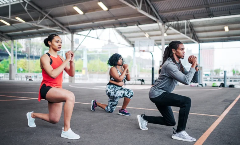
For decades, the standard protocol for preventing knee injuries in both professional sports and general fitness has centered almost exclusively on the musculature surrounding the thigh. Athletes and physical therapists have prioritized the quadriceps and hamstrings, operating under the assumption that these large muscle groups act as the primary stabilizers for the knee joint. However, emerging research from the Peak Performance Project (P3) and insights from movement specialists like Abbott are challenging this long-held convention. New data suggests that the key to mitigating the risk of Anterior Cruciate Ligament (ACL) tears and chronic knee instability lies not above the knee, but in the often-neglected muscles of the lower leg: the soleus and the posterior tibialis.
This paradigm shift in sports medicine comes at a critical time. ACL injuries remain one of the most debilitating setbacks for athletes, often requiring extensive surgery and a recovery period of nine to twelve months. By shifting the focus to the biomechanics of the foot and ankle, researchers have identified a specific movement pattern that precedes nearly every non-contact ACL injury, offering a new roadmap for preventative training that could reduce injury rates by more than two-thirds.
The Biomechanics of Injury: Identifying the Translation Pattern
The foundation of this new understanding stems from an exhaustive multi-year study conducted by the Peak Performance Project, a world-renowned sports science facility that utilizes advanced motion-capture technology and force plates to analyze athletic movement. By tracking nearly 400 National Basketball Association (NBA) players over several seasons, P3 researchers amassed millions of data points regarding landing mechanics, lateral movement, and jumping profiles.
The most significant finding of this study was the identification of a specific landing error termed "translation." Analysts discovered that every single player in the study who subsequently suffered an ACL tear exhibited the same mechanical flaw: upon landing, they touched the ground with the outside of the foot first, followed by an immediate and uncontrolled inward roll of the weight. This inward collapse forces the tibia, or shinbone, into a "windshield wiper" motion.
In a neutral, stable landing, the tibia remains relatively vertical or moves in a linear plane with the foot. However, during a translation event, the inward rolling of the foot forces the tibia to rotate and shift laterally while the femur remains relatively fixed or rotates in the opposite direction. This creates a shearing force that places the ACL under extreme tension. When the force of the landing exceeds the ligament’s tensile strength, a rupture occurs. The research indicates that this vulnerability is rarely a result of weak quads, but rather a failure of the lower leg muscles to stabilize the foot and ankle upon impact.
The Hidden Heroes: Soleus and Posterior Tibialis
To counteract the dangerous translation pattern, sports scientists are now highlighting the "under-recognized heroes" of the lower body. While the gastrocnemius—the large, visible calf muscle—receives the most attention in the gym, it is the deeper muscles that provide the necessary stability for injury prevention.
The soleus is a powerful muscle located beneath the gastrocnemius. Unlike its more famous neighbor, which crosses both the knee and ankle joints, the soleus only crosses the ankle. Its primary function is to maintain postural stability and control the forward lean of the shin during movement. Furthermore, the soleus acts as a "second heart," playing a vital role in pumping venous blood from the lower extremities back to the torso. In the context of injury prevention, a strong soleus allows an athlete to maintain a "loaded" ankle position, ensuring that the force of a landing is distributed through the Achilles tendon rather than being dumped directly into the knee joint.
The posterior tibialis is equally crucial. Located deep in the back of the shin, it is the primary stabilizing muscle for the arch of the foot. Its main role is to prevent the foot from over-pronating or rolling inward. When the posterior tibialis is weak or fatigued, the arch collapses upon landing, triggering the inward roll that leads to the aforementioned tibial translation. By strengthening these specific muscles, athletes can create a rigid, stable base that dictates how force travels upward through the kinetic chain.
A New Protocol for Force Absorption
The transition from a vulnerable landing to a resilient one involves a sequence of muscle engagements that researchers call the body’s "shock absorption system." When the lower leg muscles are properly conditioned, the mechanics of a jump or a sudden stop change fundamentally.

The ideal sequence begins with the ball of the foot making contact with the ground while the ankle is in a dorsiflexed, "loaded" state. This engagement allows the Achilles tendon—the strongest tendon in the human body—to act as a primary spring, absorbing the initial kinetic energy. From the Achilles, the force is transitioned through the lower leg to the quadriceps, and finally to the gluteus maximus, the body’s largest and most powerful muscle group.
This distribution of force across three major joints (ankle, knee, and hip) and their respective muscle groups significantly reduces the load on any single structure. In contrast, an unstable ankle forces the knee to absorb a disproportionate amount of energy in a compromised position. The P3 protocol emphasizes that injury prevention is less about the strength of a single muscle and more about the efficiency of this force-sharing system.
Data-Driven Results: A 67% Reduction in Risk
The implications of these findings are supported by remarkable statistical evidence. According to data released by Abbott and the P3 research team, athletes who implement specific lower-leg strengthening interventions see a reduction in ACL tears of up to 67%. To put this in perspective, few medical interventions or surgical procedures in sports medicine boast a success rate of this magnitude.
This 67% figure represents a potential revolution in how professional leagues manage player health. In the NBA and NFL, where a single ACL injury can cost a franchise millions of dollars in lost player productivity and contract value, the adoption of lower-leg focused "prehab" is becoming a standard requirement.
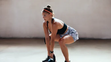
The intervention itself is notably accessible. Unlike high-intensity plyometrics or heavy powerlifting, which carry their own inherent risks, training the soleus and posterior tibialis involves targeted, controlled movements. These include seated calf raises (which isolate the soleus by keeping the knee bent) and resisted inversion exercises for the posterior tibialis. The research suggests that consistency is more important than intensity; even a few minutes of dedicated lower-leg work twice a week can "rewire" the neuromuscular pathways responsible for landing stability.
Chronology of Sports Science Evolution
The shift toward lower-leg focus is the latest chapter in a decades-long evolution of ACL research:
- 1980s-1990s: The focus was primarily on surgical techniques. ACL tears were often seen as "career-ending," and rehabilitation focused on regaining basic range of motion.
- 2000s: The "Quad-Centric" Era. Research emphasized the importance of quadriceps strength in protecting the knee, leading to the widespread use of leg extensions and squats in rehab protocols.
- 2010s: The Rise of Functional Movement. Specialists began looking at the "Valgus Collapse" (knees caving in) and started emphasizing glute medius strength to stabilize the hips.
- 2020s-Present: The Data-Driven Era. Facilities like P3 use high-speed cameras and force plates to identify that the problem often starts at the ground level. The focus moves to the foot-ankle complex and the deep stabilizers of the lower leg.
Broader Implications for Public Health and Longevity
While the initial research focused on elite NBA athletes, the findings have profound implications for the general population. Knee pain and ligamentous injuries are among the leading causes of physical inactivity in middle-aged and older adults. By applying the principles of lower-leg resilience, everyday exercisers—from weekend runners to hikers—can protect their joints against the wear and tear of daily activity.
Medical professionals are beginning to integrate these findings into geriatric care as well. Since the soleus is critical for balance and blood circulation, strengthening this muscle can reduce the risk of falls in the elderly and improve cardiovascular efficiency. The "translation" pattern identified in basketball players is often mirrored in the unsteady gait of older adults, suggesting that lower-leg stability is a fundamental requirement for mobility across the entire lifespan.
Conclusion and Future Outlook
The work of Abbott and the researchers at P3 has provided a definitive link between lower-leg mechanics and upper-leg health. By identifying the "translation" pattern and the 67% reduction in injury risk through targeted intervention, they have provided a blueprint for a more resilient human body.
As this knowledge permeates the fitness industry, the traditional "leg day" is likely to be redesigned. Future training programs will likely place as much emphasis on the soleus and posterior tibialis as they currently do on the hamstrings and glutes. For the athlete, this means more time on the court and less time in the training room. For the general public, it means the freedom to remain active, confident, and injury-free well into the later stages of life. The "hidden heroes" below the knee are finally receiving the recognition they deserve as the foundation of human movement.


