
Mitochondrial Pearling: A Newly Understood Mechanism for Organizing Cellular Powerhouses
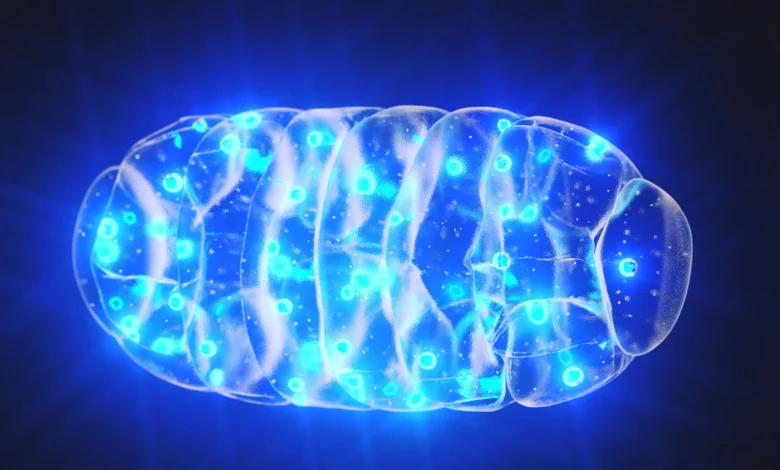
Mitochondria, the indispensable power plants of our cells, are responsible for generating the vast majority of the energy required for cellular life. This vital function is intrinsically linked to their possession of their own distinct genetic material, known as mitochondrial DNA (mtDNA). Far from being a passive bystander, mtDNA is meticulously organized within these organelles, a process scientists have long sought to understand. New research has now illuminated a previously underappreciated physical mechanism, "mitochondrial pearling," as the key to maintaining this crucial order, with profound implications for understanding cellular health and disease.
The Crucial Role of Mitochondrial DNA Organization
Within each eukaryotic cell, hundreds to thousands of copies of mtDNA reside. These copies are not scattered haphazardly but are instead organized into compact structures called nucleoids. For decades, researchers have observed a remarkably consistent, regular spacing of these nucleoids within the mitochondria. This precise arrangement is not merely an aesthetic detail; it is fundamental to the reliable transmission of mtDNA to daughter cells during division and ensures the equitable expression of the genes encoded within it across the entire mitochondrion.
The consequences of disrupted mitochondrial function, and by extension, disorganized mtDNA, are severe and far-reaching. These disruptions have been implicated in a spectrum of debilitating metabolic and neurological conditions, including liver failure and encephalopathy. Furthermore, compromised mitochondrial health is a hallmark of aging-related disorders such as Alzheimer’s and Parkinson’s disease, underscoring the critical importance of maintaining the integrity and organization of these cellular powerhouses.
A Long-Standing Enigma in Cell Biology
Despite the well-established significance of mtDNA organization, the precise mechanisms by which cells achieve and maintain this consistent nucleoid spacing have remained elusive. Previous hypotheses, focusing on dynamic processes like mitochondrial fusion and fission, or molecular tethering mechanisms, have proven insufficient to explain the observed regularity. "Proposed mechanisms related to mitochondrial fusion, fission, or molecular tethering cannot explain it, since nucleoid spacing is maintained even when they are disrupted," stated Suliana Manley, professor at the Laboratory of Experimental Biophysics (LEB) at EPFL.
This gap in knowledge presented a significant challenge to understanding the fundamental principles of cellular bioenergetics and disease pathogenesis. The precise regulation of mtDNA distribution is not a minor detail; it is a cornerstone of cellular vitality.
Unveiling the Mechanism: Mitochondrial Pearling Takes Center Stage
A groundbreaking study by Suliana Manley and her colleague Juan Landoni, a postdoctoral fellow at the LEB, has now identified the key process responsible for this long-standing mystery: mitochondrial pearling. This transient, dynamic shape change, previously given little scientific attention, involves mitochondria temporarily adopting a "beads on a string" morphology. It is during this distinctive transformation that clusters of mtDNA are effectively separated and redistributed, leading to a more even spread of nucleoids and the preservation of their regular spacing.
Observing Mitochondrial Dynamics in Real-Time
To unravel the intricacies of mitochondrial pearling, Landoni and Manley employed a sophisticated suite of advanced imaging techniques. These included super-resolution imaging, which offers unparalleled detail of cellular structures; correlated light and electron microscopy (CLEM), enabling the fusion of live-cell imaging with ultra-high-resolution electron microscopy; and phase contrast microscopy, a label-free technique that visualizes cellular structures based on differences in refractive index.
By integrating these powerful tools, the research team achieved unprecedented insights. They were able to track individual nucleoids, capture the rapid, fleeting changes in mitochondrial shape associated with pearling, and gain a clearer understanding of the internal organizational dynamics at play within these vital organelles. This multi-pronged approach provided a comprehensive view of the process, moving beyond static snapshots to dynamic, real-time observation.
The Dynamics of Mitochondrial Pearling
The live-cell imaging data revealed that mitochondrial pearling is a surprisingly frequent event, occurring multiple times per minute in actively functioning cells. During these pearling episodes, mitochondria transiently develop evenly spaced constrictions along their tubular structure, creating a series of interconnected, bulge-like segments. Crucially, the distance between these "pearls" closely mirrors the typical spacing observed between nucleoids in a non-pearling mitochondrion.
Analysis of these bead-like segments showed that most of them contain a nucleoid situated near their center. However, the study also noted that these pearling structures can form even in the absence of mtDNA, suggesting that the physical scaffolding of the mitochondrion plays a primary role in dictating this shape change.
As the pearling process unfolds, larger agglomerations of nucleoids frequently fragment into smaller groups. These dispersed nucleoids then settle into adjacent pearl-like compartments. When the mitochondrion subsequently reverts to its more common tubular form, the nucleoids remain relatively separated, thereby consolidating the even distribution that was established during the pearling phase. This cyclical process effectively acts as an internal sorting and spacing mechanism.
Unraveling the Regulatory Control of Pearling
Beyond observing the phenomenon, the researchers delved into the factors that trigger and regulate mitochondrial pearling. Through a series of meticulous genetic and pharmacological experiments, they identified key players in this process. Their findings indicate that the influx of calcium ions into the mitochondria can serve as a potent trigger for pearling. Furthermore, the internal membrane structures within the mitochondrion appear to play a crucial role in facilitating and maintaining the separation of nucleoids during these events.
Conversely, when these regulatory factors are perturbed, the researchers observed a tendency for nucleoids to aggregate rather than maintain their characteristic even spacing. This disruption of regulatory control highlights the delicate balance required for maintaining mitochondrial genome integrity and function. The ability to manipulate these regulatory factors opens avenues for therapeutic interventions in conditions where such aggregation is detrimental.
A Rediscovered Historical Observation
The significance of mitochondrial pearling extends beyond its newly elucidated functional role. Juan Landoni noted the historical context of the discovery: "Since Margaret Reed Lewis first sketched mitochondrial pearling in 1915, it has largely been dismissed as an anomaly linked to cellular stress." For over a century, this intriguing morphological change was relegated to the footnotes of cell biology, often considered an artifact of experimental conditions or cellular distress rather than a fundamental cellular process.
"Over a century later, it is emerging as an elegantly conserved mechanism at the heart of mitochondrial biology," Landoni continued. "This biophysical process offers a simple and energy efficient means to distribute the mitochondrial genome." The rediscovery and functional validation of Lewis’s early observations underscore the enduring value of meticulous scientific observation and the potential for seemingly minor details to hold profound biological significance.
Broader Implications for Health and Disease
This pivotal discovery that cells rely on physical processes, not solely complex molecular machinery, for organization has far-reaching implications. Understanding the precise workings of mitochondrial pearling and the intricate network of factors that control it could unlock crucial insights into a wide range of diseases linked to mtDNA dysfunction.
The implications for therapeutic development are particularly significant. By elucidating the natural mechanisms that maintain mitochondrial genome order, researchers may be able to devise novel strategies to correct or prevent the aberrant nucleoid clumping seen in various pathologies. This could lead to new treatment approaches for neurodegenerative diseases, metabolic disorders, and age-related conditions where mitochondrial health is compromised. For example, identifying specific molecular targets that can promote or enhance pearling in situations of stress or disease could offer a powerful new therapeutic avenue. The development of drugs that modulate calcium influx or influence internal mitochondrial membrane dynamics could become a focus of future pharmaceutical research aimed at combating mitochondrial dysfunction.
The Path Forward: Research and Collaboration
The work by Manley and Landoni represents a significant leap forward in our understanding of cellular organization and bioenergetics. Their findings not only solve a long-standing mystery but also open new avenues for research into the fundamental processes that govern cellular life and health. Future research will likely focus on further dissecting the molecular players involved in regulating pearling, exploring its role in different cell types and under various physiological conditions, and investigating its potential as a therapeutic target.
The collaborative efforts of scientists across disciplines, from biophysics and cell biology to genetics and pharmacology, will be essential in fully realizing the potential of this discovery. The journey from a historical sketch to a fundamental understanding of a cellular mechanism highlights the iterative and often surprising nature of scientific progress, promising a brighter future for the treatment of diseases rooted in cellular dysfunction.


