
Texas A&M Researchers Pioneer Customizable Vessel-Chip Technology to Replicate Complex Human Vascular Architectures and Advance Disease Modeling
The human vascular system is an intricate network of biological engineering, characterized by a labyrinth of vessels that bend, branch, taper, and expand to facilitate the transport of life-sustaining blood. For decades, the scientific community has struggled to replicate this complexity in a laboratory setting, often relying on simplified models that treated blood vessels as uniform, straight tubes. While these rudimentary designs provided a baseline for understanding basic fluid dynamics, they failed to capture the physiological nuances where the most critical vascular diseases—such as atherosclerosis, aneurysms, and thrombosis—typically originate. Addressing this technological gap, researchers at the Department of Biomedical Engineering at Texas A&M University have developed a groundbreaking, customizable "vessel-chip" system designed to mirror the authentic structural diversity of human anatomy.
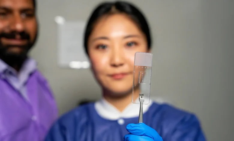
This innovative platform, emerging from the Bioinspired Translational Microsystems Laboratory, represents a significant leap forward in the field of organs-on-a-chip (OOC). By allowing scientists to recreate the specific geometries of patient-specific vasculature, the system provides a more realistic environment for studying disease progression and testing the efficacy of new pharmacological interventions. The research, led by Associate Professor Dr. Abhishek Jain and master’s student Jennifer Lee, marks a transition from static, idealized models to dynamic, "living" systems that can be tailored to the unique physiological profiles of individual patients.
Engineering Complexity: Beyond the Straight Tube
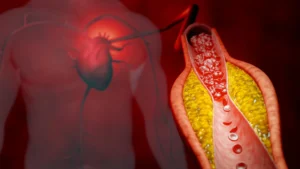
The fundamental limitation of traditional vascular models lies in their inability to simulate the mechanical forces generated by complex blood flow. In a healthy human body, blood does not move through a series of perfect cylinders; instead, it navigates a series of bifurcations and varying diameters. These structural variations dictate the "shear stress"—the frictional force exerted by flowing blood on the inner lining of the vessel. When vessels narrow (stenosis) or bulge (aneurysms), these flow patterns change drastically, often triggering cellular responses that lead to inflammation or clot formation.
Jennifer Lee, the primary researcher on the project, emphasized that the goal was to capture these specific anomalies. "There are branched vessels, or aneurysms that have sudden expansion, and then stenosis that restricts the vessel," Lee explained. "All these different types of vessels cause the blood flow pattern to be significantly changed, and the inside of the blood vessel is affected by the level of shear stress caused by these flow patterns. That’s what we wanted to model."
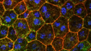
The vessel-chip itself is a microfluidic device—a specialized piece of hardware that manipulates minute amounts of fluids through channels at a microscopic scale. By using advanced manufacturing techniques, the Texas A&M team can now "print" or mold these channels into shapes that precisely mimic a patient’s own vascular imaging. Once the architecture is established, the researchers coat the interior of the channels with human endothelial cells—the cells that form the interface between circulating blood and the vessel wall. This creates a "living" model that responds to fluid pressure and chemical signals just as a real vessel would.
A Chronology of Innovation at Texas A&M
The development of the customizable vessel-chip is the result of years of iterative research within the Bioinspired Translational Microsystems Laboratory. The journey toward this May 2025 cover-story publication in the journal Lab on a Chip began with foundational work by Dr. Tanmay Mathur, a former graduate student under Dr. Jain’s mentorship.
Several years ago, Dr. Mathur developed the lab’s first-generation straight vessel-chip. This early model was instrumental in proving that microfluidic devices could support living endothelial cells under flow conditions. However, the team recognized that the "straight-tube" approach was an oversimplification. When Jennifer Lee joined the lab as an undergraduate honors student, she was tasked with evolving this design into a more sophisticated platform capable of handling non-linear geometries.
Lee’s progression from an undergraduate researcher to a Master of Science student through Texas A&M’s fast-track program allowed for a continuity of research that is rare in academic settings. Dr. Jain noted that the fast-track program was essential for taking "high-impact, high-risk" research from a conceptual stage to a published outcome. This timeline of development—moving from basic straight-line flow to complex, multi-dimensional architecture—reflects a broader trend in biomedical engineering toward increasing biological fidelity.
The Science of Shear Stress and Disease Localization
To understand the impact of this technology, one must look at the data regarding where vascular diseases occur. Clinical data shows that atherosclerotic plaques do not form randomly throughout the circulatory system; they predominantly develop at "sites of predilection," such as the curvatures of the aorta or the branching points of the carotid arteries. At these locations, blood flow becomes turbulent or "disturbed," leading to low or oscillatory shear stress.
By utilizing the customizable vessel-chip, the Texas A&M team can now recreate these exact conditions in a controlled environment. This allows for the observation of how endothelial cells change their gene expression and physical behavior in response to different shapes. For instance, in a modeled "stenosis" (a narrowed vessel), the researchers can measure the high-velocity flow and the resulting high shear stress that can lead to platelet activation and heart attacks. Conversely, in an "aneurysm" model, they can study the stagnant flow areas that are prone to the development of blood clots.
"We can now start learning about vascular disease in ways we’ve never been able to before," said Dr. Jain. "Not only can you make these structures complex, you can put actual cellular and tissue material inside them and make them living. These are the sites where vascular diseases tend to develop, so understanding them is critical."
Advancing the "Fourth Dimension" of Organ-on-a-Chip
While the current iteration of the vessel-chip focuses on the interaction between flow and endothelial cells, the research team is already looking toward the next frontier. Dr. Jain describes this as the "fourth dimensionality" of organs-on-a-chip. This concept moves beyond three-dimensional structure and adds the element of complex biological interaction over time.
Future versions of the chip are expected to incorporate additional cell types, such as smooth muscle cells, pericytes, and even immune cells like macrophages. In a real human vessel, the endothelial lining communicates constantly with the underlying muscle layers. By adding these layers to the chip, researchers can study how a signal from the blood flow penetrates through the vessel wall, potentially leading to better treatments for hypertension and chronic inflammatory diseases.
This multi-cellular approach is particularly relevant for drug testing. Currently, the pharmaceutical industry relies heavily on animal models, which often fail to accurately predict human responses due to species-specific differences in vascular biology. The vessel-chip offers a "human-on-a-chip" alternative that could significantly reduce the time and cost of drug development while improving safety profiles.
Supporting Data and Institutional Backing
The significance of this research is underscored by the high level of institutional and federal support it has received. The project is a collaborative effort funded by a consortium of the United States’ most prominent scientific and regulatory bodies. Supporting organizations include:
- The National Institutes of Health (NIH) and the U.S. Food and Drug Administration (FDA): Interested in the chip’s potential to provide more accurate data for regulatory drug approval.
- NASA: Investigating how microgravity and space radiation affect the vascular health of astronauts, where vessel-chips can serve as portable, lightweight testing platforms.
- The U.S. Army Medical Research Program: Focusing on trauma-induced vascular injuries and rapid treatment protocols.
- The Biomedical Advanced Research and Development Authority (BARDA): Looking at the vascular effects of chemical or biological threats.
This broad spectrum of funding highlights the versatile applications of the vessel-chip, ranging from deep-space exploration to domestic public health.
Implications for Future Medical Research
The implications of customizable vessel-chips extend far beyond the laboratory. In the realm of personalized medicine, a clinician could theoretically take a biopsy of a patient’s cells, grow them within a vessel-chip modeled after the patient’s own unique vascular anatomy (obtained via MRI or CT scan), and test various medications to see which one most effectively prevents clotting or inflammation for that specific individual.
Furthermore, the technology addresses a growing ethical and practical demand to reduce animal testing. As the pharmaceutical industry faces increasing pressure to move away from animal models, "organ-on-a-chip" systems provide a scientifically superior alternative that uses human cells to predict human outcomes.
Beyond the technical achievements, the project has also served as a catalyst for professional development. Jennifer Lee credits the collaborative environment of the Bioinspired Translational Microsystems Laboratory with providing her with skills in problem-solving and communication that are vital for the next generation of engineers. "It’s such a good environment to interact with not only peers but also graduate students and postdoctoral researchers," Lee said. "You’re able to learn teamwork and communication, work ethic, and just trying different things out."
As the Texas A&M team prepares for the May 2025 cover feature in Lab on a Chip, the focus remains on refining the complexity of these models. The transition from simple tubes to living, breathing, and branching replicas of human anatomy marks a new era in biomedical science—one where the mysteries of the human heart and its vast network of vessels are closer than ever to being solved.

