The seemingly innocuous act of using a public restroom can, for many, be a source of considerable anxiety. Parents of young children, individuals managing chronic health conditions that necessitate frequent bathroom visits, and indeed, a significant portion of the general population, have developed an intuitive, and often learned, understanding of which public facilities are to be approached with caution and which are best avoided altogether. This practical knowledge, honed through experience, often dictates our choices in moments of urgent need. However, even with this awareness, there are unavoidable instances where one must utilize a facility that appears far from pristine. In such situations, a common dilemma arises: is it truly safe to sit down, even if the toilet seat presents a passable appearance? Could such a decision lead to illness? This exploration delves into the hidden world of public toilet hygiene, the science behind germ transmission, and practical strategies for mitigating risk.
The Microbial Menagerie Within Public Toilets
The human body is a complex ecosystem, and its daily output is a significant contributor to the microbial environment of toilets. On average, an adult excretes over a liter of urine and more than 100 grams of fecal matter daily. This waste is not merely inert matter; it carries with it a diverse array of bacteria and viruses that are shed from the body. These microbes, originating from the gastrointestinal tract and other bodily systems, inevitably find their way into the toilet bowl.
The situation is exacerbated for individuals experiencing conditions like diarrhea. During such episodes, the body can expel significantly higher quantities of potentially harmful microbes. In public restrooms, particularly those with high foot traffic and infrequent cleaning schedules, these organisms can rapidly accumulate. Researchers have aptly described such environments as a "microbial soup," a complex mixture of bacteria, viruses, and other microorganisms suspended in moisture and on surfaces.
Identifying the Culprits: Common Germs on Toilet Surfaces
Numerous scientific studies have been conducted to identify the types of microorganisms that inhabit public toilet surfaces. These investigations have revealed a broad spectrum of microbial life. While specific findings can vary based on geographic location, user demographics, and cleaning protocols, common culprits identified include:
- Escherichia coli (E. coli): A common bacterium found in the intestines of humans and animals. Certain strains can cause severe food poisoning, while others are harmless.
- Staphylococcus aureus: This bacterium can cause a range of infections, from skin infections to more serious conditions like pneumonia and bloodstream infections.
- Norovirus: A highly contagious virus that causes vomiting and diarrhea, often referred to as the "stomach flu."
- Shigella: Bacteria that can cause dysentery, a severe form of diarrhea often accompanied by fever and abdominal cramps.
- Salmonella: A group of bacteria commonly associated with food poisoning, causing fever, diarrhea, and abdominal cramps.
- Coliform Bacteria: A broad group of bacteria that indicates fecal contamination. Their presence suggests that other, potentially more harmful, pathogens may also be present.
Beyond individual microbes, a significant concern is the presence of biofilm. This is not a single organism but rather a complex, slimy layer formed by a community of microorganisms that adhere to surfaces. Biofilms can develop under the toilet rim, on the exterior of the bowl, and across various surfaces within the restroom. These layers can act as protective shields for bacteria, making them more resistant to cleaning agents and disinfectants. The sticky nature of biofilm also facilitates the adhesion and survival of pathogens.
Beyond the Seat: The True Hotspots of Contamination
In the common perception, the toilet seat is often considered the dirtiest part of a public restroom. However, scientific research consistently challenges this notion. Studies have demonstrated that toilet seats frequently harbor fewer microbes compared to other high-touch areas within the same facility.
The real germ reservoirs tend to be surfaces that are touched repeatedly throughout the day, often by hands that have not been recently washed. These include:
- Door Handles and Knobs: Both the exterior and interior door handles, as well as the mechanisms for opening and closing stalls, are prime breeding grounds for germs.
- Faucet Knobs and Handles: Turning on and off the water to wash hands is a critical juncture where cross-contamination can occur.
- Flush Levers and Buttons: The act of flushing the toilet, a necessary step after use, involves direct contact with these mechanisms.
- Dispenser Buttons (Soap, Paper Towel): Similarly, interacting with dispensers for hygiene products can transfer microbes.
In high-traffic locations such as airports, train stations, shopping malls, and public parks, restrooms can be used hundreds, or even thousands, of times per week. While some of these facilities may benefit from rigorous and frequent cleaning schedules, others, particularly those in less maintained public spaces like bus stops or remote park facilities, might only receive attention once a day or even less frequently. Visible indicators of inadequate cleaning often include persistent urine odors, dirty floors with visible grime and debris, and soiled toilet bowls.
The Unseen Threat: Toilet Plumes and Aerosolized Germs
While direct contact with surfaces is a primary mode of germ transmission, a more insidious threat lurks within the mechanics of flushing: the "toilet plume." When a toilet is flushed, especially if the lid is not closed, the force of the water can create an aerosolized cloud of tiny droplets. These droplets, propelled upwards and outwards, can carry bacteria and viruses from the toilet bowl into the surrounding air. Research indicates that these plumes can travel distances of up to 2 meters, potentially contaminating nearby surfaces and individuals.
Another factor contributing to germ dispersal is the widespread use of hot air hand dryers. While intended to promote hygiene, if hands are not thoroughly washed and dried, these devices can inadvertently blow residual microbes onto the user’s skin, onto other individuals in the vicinity, and onto surrounding surfaces. This can create a secondary wave of contamination within the restroom environment.
Pathways of Transmission: How Germs Enter Your Body
Germs in public bathrooms can enter your body through several pathways, highlighting the importance of a multi-faceted approach to personal hygiene. These include:
- Direct Contact: Touching contaminated surfaces (door handles, faucet knobs, toilet seats) and then touching your face, eyes, nose, or mouth.
- Indirect Contact: Using contaminated objects, such as your mobile phone, in the restroom and then touching your face.
- Inhalation: Breathing in aerosolized droplets from toilet plumes or from contaminated air expelled by hand dryers.
- Ingestion: Accidentally ingesting germs through contaminated food or drink consumed after using a public restroom without proper hand hygiene.
Proactive Protection: Practical Strategies to Minimize Risk
Fortunately, the risk of contracting an illness from a public restroom can be significantly reduced through the adoption of simple yet effective hygiene practices. These habits, when consistently applied, create a robust barrier against potential pathogens.
1. The Art of Handwashing: This remains the single most critical defense.
- Duration: Wash your hands thoroughly with soap and water for at least 20 seconds. This is roughly the time it takes to sing "Happy Birthday" twice.
- Technique: Lather all surfaces of your hands, including between your fingers and under your nails.
- Rinsing: Rinse thoroughly with clean, running water.
- Drying: Use clean paper towels to dry your hands completely. Avoid sharing cloth towels.
2. The Paper Towel Advantage: Opt for paper towels over air dryers whenever possible. Paper towels effectively absorb moisture and can also be used to turn off faucets and open doors, minimizing direct contact with potentially contaminated surfaces.
3. Surface Sanitization: If you are particularly concerned or if a surface appears visibly soiled, a quick wipe with an antiseptic wipe or hand sanitizer can provide an extra layer of protection for the toilet seat or other high-touch areas.
4. Personal Item Prudence: Be mindful of where you place personal items, especially mobile phones, which are notorious carriers of germs. Avoid placing them on toilet seats or sinks. Consider using a dedicated pouch or keeping them in your pocket or bag.
5. Strategic Flushing: When possible, close the toilet lid before flushing to minimize the spread of toilet plumes.
6. Hand Sanitizer as a Backup: While not a substitute for thorough handwashing, an alcohol-based hand sanitizer (with at least 60% alcohol) can be a useful alternative when soap and water are not immediately available.
7. Avoid Hovering: A common practice, particularly among those hesitant to sit, is "hovering" over the toilet. This position, while seemingly a way to avoid contact, can actually lead to other issues. It can strain the pelvic floor muscles, making it more difficult to fully empty the bladder and increasing the likelihood of splashing.
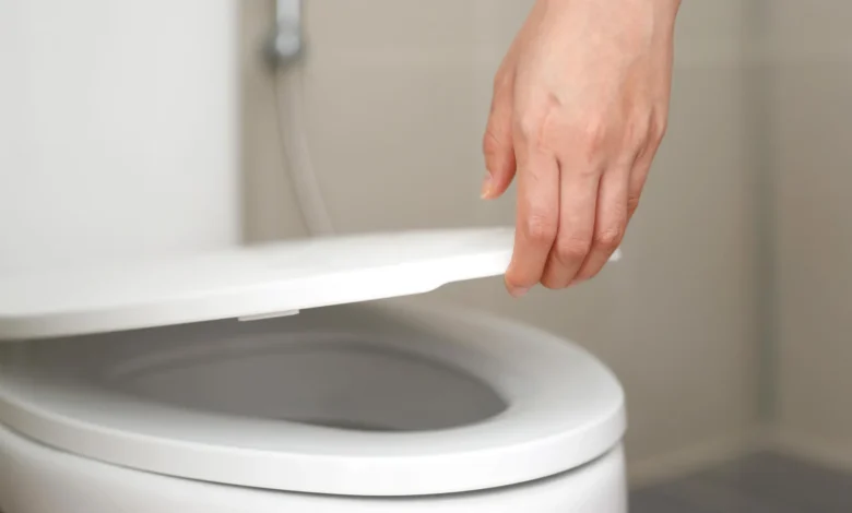
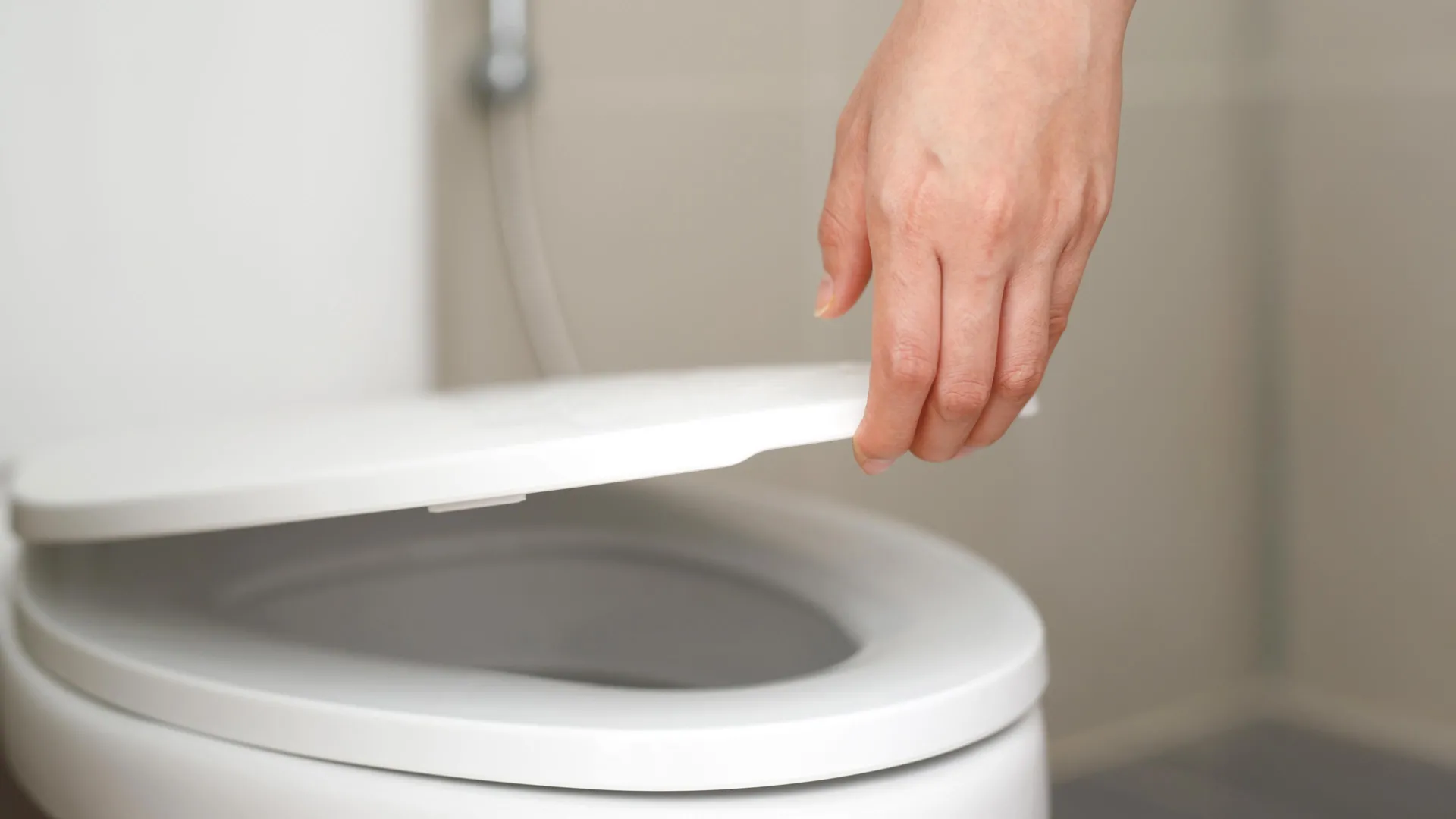
The Verdict: Is Sitting on Public Toilet Seats Safe?
For the vast majority of healthy individuals, the act of sitting on a public toilet seat poses a relatively low risk of infection. The primary concern is not the seat itself, but rather the subsequent actions and the overall hygiene practices. If you are concerned about cleanliness, a simple wipe of the seat with toilet paper or a disinfectant wipe, or the use of a disposable seat cover, can provide significant peace of mind and an additional layer of reassurance.
The infections we are more likely to acquire in public restrooms tend to stem from:
- Contaminated Hands: The most prevalent route of transmission.
- Frequently Touched Surfaces: Door handles, faucets, and flush levers.
- Airborne Droplets: From toilet plumes or inefficient hand dryers.
- Personal Items: Such as mobile phones that are used in the restroom environment.
Therefore, the most effective strategy for safeguarding your health in public restrooms is not solely focused on the toilet seat, but rather on a holistic approach to personal hygiene. Prioritizing proper handwashing, utilizing paper towels for drying, cleaning surfaces when necessary, and maintaining the cleanliness of personal items like your phone are paramount. By understanding the dynamics of germ transmission and adopting these practical, evidence-based habits, individuals can navigate public restrooms with greater confidence and significantly reduce their risk of illness. The key lies in informed action and consistent adherence to good hygiene practices, transforming a potentially concerning experience into a manageable part of daily life.


