
SPECT-CT Imaging as a Critical Diagnostic Tool in Managing Bone Metastasis for Cancer Patients

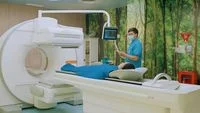
In the complex landscape of oncology, bone metastasis represents one of the most challenging aspects of cancer progression. When primary malignancies—most notably breast, prostate, and lung cancers—spread to the skeletal system, the implications for a patient’s quality of life and long-term prognosis are profound. Because bone metastasis often presents without initial, overt symptoms, early and precise detection is paramount. Modern diagnostic medicine has turned to hybrid imaging technologies, specifically the Single-Photon Emission Computed Tomography (SPECT) combined with Computed Tomography (CT), to bridge the gap between functional physiological data and high-resolution anatomical imaging.
As medical institutions seek to improve survivability and patient comfort, the integration of SPECT-CT has emerged as a cornerstone of comprehensive cancer care. By providing a holistic view of the skeletal structure, this diagnostic approach enables clinicians to identify abnormal cellular activity long before structural damage becomes irreversible.
The Mechanism and Clinical Utility of SPECT-CT
To understand why SPECT-CT has become a gold standard in bone metastasis evaluation, one must first understand the limitations of conventional imaging. Standard X-rays or basic bone scans often lack the specificity to distinguish between benign bone lesions—such as fractures from minor trauma or degenerative changes—and malignant deposits.
A SPECT scan operates by detecting gamma rays emitted by a radiopharmaceutical tracer injected into the patient. This tracer accumulates in areas of increased bone turnover, which is characteristic of metastatic activity. However, a standalone SPECT image provides poor anatomical detail; it identifies a "hot spot" without clearly showing the surrounding tissue architecture. This is where the integration of CT proves transformative. The CT scan provides the precise spatial resolution required to pinpoint exactly where the abnormal activity is occurring within the skeletal frame.
By fusing these two modalities, the SPECT-CT scan creates a multi-dimensional map of the body. This allows oncologists and nuclear medicine specialists to differentiate between metabolic activity and structural deformity, significantly reducing the rate of false positives and ambiguity in diagnostic reporting.
Clinical Perspectives on Diagnostic Precision
The role of nuclear medicine specialists in the diagnostic pathway is critical to the multidisciplinary approach favored by modern oncology centers. Dr. Yustia Tuti, a specialist in Nuclear Medicine and Molecular Imaging at Mayapada Hospital Jakarta Selatan, emphasizes that the goal is not merely to detect the presence of cancer, but to map its systemic impact.
"In managing patients with bone metastasis, the clinical focus must extend beyond the simple binary of identifying a lesion," explains Dr. Yustia. "We must evaluate the location, the spatial extent of the disease, and the current structural integrity of the bone. This information dictates whether a patient is at immediate risk for debilitating complications, such as pathological fractures or spinal cord compression, which can severely compromise patient mobility and neurological function."
The risk of these complications is not merely academic; it is a primary driver of cancer-related morbidity. Pathological fractures, occurring when a tumor weakens the bone to the point that it breaks under normal stress, often require surgical intervention and prolonged rehabilitation. Similarly, spinal cord compression is a medical emergency that necessitates immediate action to prevent permanent paralysis. By utilizing SPECT-CT, clinicians can preemptively identify bones at high risk of fracture, allowing for prophylactic stabilization or targeted radiation therapy.
Refining the Differential Diagnosis
A significant challenge in oncology is distinguishing between treatment-related changes and disease progression. Chemotherapy, radiation, and hormonal therapies can all influence bone metabolism, sometimes creating "flares" on imaging that can be misinterpreted as tumor growth.
Dr. Andreas Lim, also a specialist in Nuclear Medicine at Mayapada Hospital, highlights how SPECT-CT provides the clarity needed to avoid unnecessary changes to a patient’s treatment regimen. "The ability to distinguish between metastasis and other benign bone changes is essential for maintaining the integrity of the patient’s treatment plan," says Dr. Lim. "When results remain ambiguous, it creates anxiety for the patient and uncertainty for the care team. SPECT-CT clears this uncertainty, ensuring that subsequent medical decisions are grounded in the most accurate clinical evidence possible."
This analytical precision is essential for "precision medicine," a philosophy that seeks to tailor therapy to the individual characteristics of the patient’s disease. By confirming the exact status of the skeletal system, clinicians can determine whether to escalate systemic therapy, initiate bone-modifying agents, or shift the focus toward palliative care to maximize the patient’s quality of life.
The Evolution of Comprehensive Cancer Care
The deployment of advanced diagnostic technology like SPECT-CT is best understood within the broader context of the "Comprehensive Cancer Care" model. In this framework, the diagnostic tool is only one component of a larger ecosystem of care. Modern oncology centers are increasingly moving away from isolated specialist consultations toward a "Tumor Board" approach.
A Tumor Board typically consists of a multidisciplinary team—including medical oncologists, radiation oncologists, surgeons, radiologists, and nuclear medicine physicians—who collectively review a patient’s diagnostics to formulate a consensus treatment plan. This collaborative environment ensures that the high-resolution data provided by a SPECT-CT scan is interpreted through multiple clinical lenses, resulting in a more robust and personalized treatment strategy.
Furthermore, the patient experience is increasingly supported by "Patient Navigators." These professionals act as liaisons, helping patients navigate the complexities of their diagnosis, treatment appointments, and post-procedural care. This supportive infrastructure is designed to reduce the administrative and emotional burden on patients, allowing them to focus on recovery.
Technological Accessibility and Future Implications
As the incidence of cancer continues to rise globally, the burden on healthcare systems to provide accessible, high-quality diagnostic services has never been greater. The availability of SPECT-CT within domestic medical facilities, such as the Poli Kedokteran Nuklir at Mayapada Hospital, represents a significant leap forward in regional healthcare equity. By localizing this technology, patients are spared the logistical challenges of seeking high-level diagnostic services abroad.
Beyond the immediate clinical benefits, the data generated by these imaging platforms contributes to the broader field of medical research. As these facilities aggregate data on bone metastasis patterns, they contribute to the global understanding of cancer behavior. This continuous feedback loop—where diagnostic findings inform clinical practice, and clinical practice informs future research—is essential for improving long-term survival rates.
Supporting the Patient Journey: Data and Connectivity
In addition to diagnostic imaging, the integration of digital health tools is becoming a standard feature of modern healthcare. Applications such as "MyCare" offer patients a mechanism for self-monitoring, tracking vital indicators like heart rate, activity levels, and BMI. While these metrics do not replace clinical diagnostics, they empower patients to maintain an active role in their general health, providing a supplementary layer of information for doctors to evaluate during routine check-ups.
The combination of high-tech imaging and patient-centric digital tools reflects a shift toward a more holistic view of the cancer patient. Treatment is no longer just about attacking the tumor; it is about sustaining the patient’s physical and emotional well-being throughout the duration of the disease.
Summary of Diagnostic Impact
The clinical adoption of SPECT-CT has fundamentally changed the evaluation of bone metastasis. By providing superior spatial resolution and functional data, it allows for:
- Early Detection: Identifying metastatic lesions before they manifest as chronic pain or physical disability.
- Risk Stratification: Assessing the structural stability of the bone to predict and prevent pathological fractures.
- Treatment Monitoring: Accurately assessing how a patient is responding to systemic therapy and differentiating between healing bone and active disease.
- Multidisciplinary Alignment: Providing a common, high-quality data set that allows diverse medical specialists to align on a singular treatment goal.
As oncology continues to evolve, the integration of such sophisticated imaging technologies will remain vital. For patients facing the uncertainty of cancer, the clarity provided by these diagnostic advancements is not just a medical milestone; it is a critical component of the path toward better health outcomes. For those requiring further consultation, institutional resources such as the Mayapada Hospital Call Center (150770) and specialized digital services serve as the necessary bridges to accessing these life-saving diagnostics.
Ultimately, the goal remains clear: to transition from reactive treatment to proactive, evidence-based care, ensuring that every patient receives the most precise evaluation possible, tailored specifically to the unique contours of their own anatomy and disease progression.