Cellular Limbo: How Alcohol-Induced Inflammation Traps Liver Cells and Halts the Body’s Ultimate Regenerative Engine

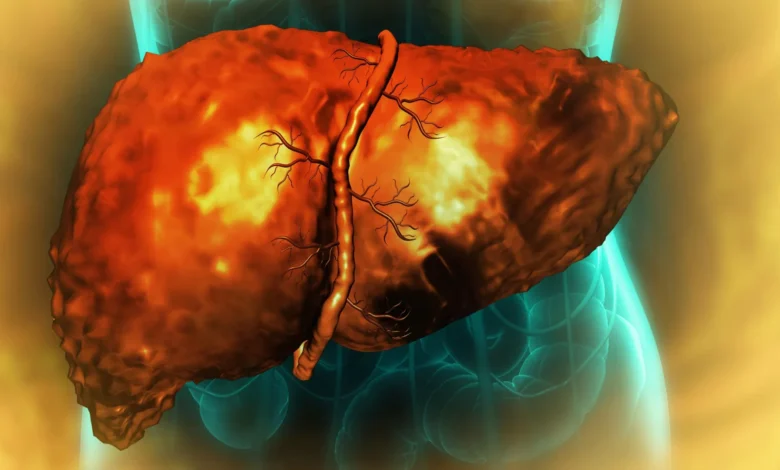
The human liver is widely recognized as one of the body’s most resilient internal organs, possessing a near-miraculous capacity to repair, rebuild, and restore itself even after sustaining substantial injuries or undergoing partial surgical removal. However, a groundbreaking collaborative study reveals that chronic, excessive alcohol consumption can fundamentally disable this vital biological machinery. According to new research published in the journal Nature Communications, alcohol-related liver damage leaves critical liver cells trapped in an abnormal, unproductive "middle state." These cells become perpetually incapable of functioning normally or completing the regeneration cycle, remaining paralyzed even long after an individual has completely stopped drinking.
This cellular entrapment explains why patients suffering from advanced alcohol-associated liver disease (AALD) often experience progressive, irreversible liver failure. Conducted by an interdisciplinary team of researchers from the University of Illinois Urbana-Champaign, Duke University, and the Chan Zuckerberg Biohub Chicago, the study sheds critical light on the molecular mechanisms of organ failure and opens promising new avenues for targeted diagnostics and future pharmacological interventions.
The Global Toll of Alcohol-Associated Liver Disease
To understand the magnitude of this discovery, medical experts point to the staggering global burden of alcohol-associated liver disease. AALD stands as the leading cause of liver-related mortality worldwide, accounting for roughly three million deaths annually. The spectrum of the disease ranges from early-stage fatty liver and alcoholic hepatitis to advanced cirrhosis, a condition characterized by extensive scarring and loss of organ architecture.
Under normal physiological conditions, the liver manages minor injuries through a sophisticated cellular balancing act. Surviving hepatocytes—the primary functional cells of the liver—temporarily alter their identity, de-differentiating into a flexible, fetal-like progenitor state. These progenitor cells proliferate rapidly to replace lost or damaged tissue, and subsequently re-differentiate back into mature, fully functional adult cells once tissue mass is restored.
Yet, in patients suffering from severe AALD, this elegant cycle breaks down entirely. Clinicians and researchers have long been perplexed by a clinical reality: why do patients with severe alcoholic hepatitis and cirrhosis continue to deteriorate and experience end-stage liver failure even after achieving complete sobriety?
"We knew that the liver stops functioning and stops regenerating in patients with alcohol-related hepatitis and cirrhosis, even when a patient has discontinued consuming alcohol, but we didn’t know why," explained Dr. Auinash Kalsotra, a biochemistry professor at the University of Illinois Urbana-Champaign who co-led the study alongside Duke University School of Medicine professor Dr. Anna Mae Diehl. "The only real life-saving treatment option once a patient reaches the liver failure stage in those diseases is transplantation. But if we understood why these livers were failing, maybe we could intervene."
A Multicenter Quest to Unravel the Molecular Mystery
To investigate this cellular breakdown, Kalsotra and Diehl leveraged years of prior foundational research into the molecular programs governing liver regeneration. Collaborating with researchers at Johns Hopkins University School of Medicine—who provided vital human liver tissue samples through an initiative supported by the National Institute on Alcohol Abuse and Alcoholism (NIAAA), a division of the National Institutes of Health (NIH)—the team embarked on a comparative genetic and molecular analysis.
The researchers compared healthy, undamaged human liver tissue with diseased tissue samples obtained from patients diagnosed with advanced alcohol-associated hepatitis and cirrhosis. Using advanced high-throughput sequencing and computational models, the team tracked the transcriptional and post-transcriptional behavior of cells within the diseased organs.
A striking, uniform pattern quickly emerged from the data. Rather than successfully cycling between mature adult states and proliferating progenitor states, cells in the damaged livers had initiated the transition toward regeneration but had become permanently stalled halfway.
"They are neither functional adult cells nor proliferative progenitor cells," noted University of Illinois graduate students Ullas Chembazhi and Sushant Bangru, who served as co-first authors of the study. "Since they are not functioning, more pressure builds on the remaining cells. So they try to regenerate, and they’re all ending up in this unproductive quasi-progenitor state, and that’s what is causing liver failure."
This creates a vicious physiological feedback loop. As accumulating cells become trapped in this unproductive limbo, the total pool of functional hepatocytes shrinks. The remaining healthy cells, forced to shoulder the entire metabolic and detoxification workload of the body, experience elevated stress. Driven by this escalating functional deficit, these surviving cells attempt to regenerate, only to succumb to the same cellular trap. The organ spirals into a state of cascading failure.
RNA Splicing and the ESRP2 Protein Deficit
To pinpoint the exact biological mechanism halting cell maturation, the research team looked beyond mere gene expression counts and examined RNA processing. RNA serves as the indispensable intermediary translating genetic instructions stored in DNA into functional proteins. Before messenger RNA (mRNA) can be translated into proteins, intervening segments must be precisely excised and rejoined in a critical editing process known as RNA splicing.
RNA splicing is paramount to cellular diversity and function, allowing a single gene to produce multiple protein variants tailored to specific physiological needs. Utilizing deep RNA sequencing, the team discovered that RNA was being mis-spliced on a massive scale throughout alcohol-damaged livers, impacting thousands of distinct genes and disrupting core protein functions.
Further investigation revealed a primary driver behind these widespread splicing errors: a severe deficiency in a specialized RNA-binding protein known as ESRP2. Under normal circumstances, ESRP2 binds to RNA molecules to ensure they are edited and spliced accurately. In the alcohol-damaged human liver samples, ESRP2 levels were profoundly depleted.
The consequences of this ESRP2 deficit extended beyond protein production failures. In many instances, the mis-spliced RNA altered the molecular localization signals—the internal "zip codes" that direct synthesized proteins to their correct destinations within the cell.
"Proteins function at a very specific place in the cell, and that is directed by sequences within the protein that take the protein to that particular spot," Kalsotra explained. "We found that, in many cases, the sequence that dictates where the protein localizes within a cell was misspliced. That’s why it was important that we did the multiple analyses we did. There was the same amount of RNA and protein, but the protein was not at the right place to function. Due to missplicing, key proteins that are required for productive liver regeneration were getting stuck in the cytoplasm, when they needed to be in the nucleus."
Because the cell’s DNA resides within the nucleus, vital transcription factors and regenerative proteins trapped in the surrounding cytoplasm remain functionally idle, unable to switch on the genes required for tissue repair.
Inflammation as the Ultimate Culprit
To confirm that the loss of ESRP2 directly caused the regeneration failure, the research team conducted translational experiments using murine (mouse) models. Genetically engineered mice lacking the gene responsible for producing ESRP2 developed liver injuries and regenerative blocks that closely mirrored the pathology observed in human patients with advanced AALD.
This finding prompted a critical follow-up question: What triggers the depletion of ESRP2 in the first place?
The study points to chronic inflammation as the primary catalyst. When the liver metabolizes excess alcohol, the chemical stress damages local tissue, triggering an influx of immune cells and hepatic stellate cells to the injury sites. These activated immune and support cells release a flood of inflammatory cytokines and growth factors into the microenvironment.
The researchers discovered that these inflammatory signaling molecules actively suppress both the synthesis and functional activity of ESRP2.
To test whether this inflammatory cascade could be interrupted, the team conducted in vitro experiments using laboratory cultures of liver cells. By introducing a blocking molecule targeted against the receptor of a key inflammation-promoting factor, the researchers observed a remarkable reversal: ESRP2 levels recovered, and RNA splicing patterns returned toward normal parameters.
Implications for Future Therapeutics and Diagnostics
The identification of this inflammatory-splicing axis provides a novel conceptual framework for treating alcohol-associated liver disease. Historically, therapeutic strategies have focused either on lifestyle modifications—such as achieving sobriety—or end-stage interventions like liver transplantation, which remain severely constrained by donor organ shortages.
By demonstrating that alcohol-induced inflammation directly disrupts RNA splicing via ESRP2 suppression, the study highlights potential new targets for drug development. Rather than attempting to directly manipulate or replace failing liver tissue, future pharmacological therapies could focus on neutralizing specific inflammatory pathways to restore normal RNA processing and unlock the liver’s innate regenerative capacity.
Furthermore, the researchers suggest that the abnormally spliced RNA molecules identified in the study could serve as valuable non-invasive biomarkers. Detecting these mis-spliced RNAs in patient blood or tissue samples could enable earlier diagnosis, more accurate disease staging, and real-time monitoring of therapeutic responses.
"I’m hopeful these findings will become a launching pad for future clinical studies," Kalsotra stated. "We can use these mis-spliced RNAs as diagnostic markers or develop treatments that can curb the inflammation. And if we can correct the splicing defects, then maybe we can improve recovery and restore damaged livers."
Collaboration and Financial Support
The multi-institutional study brought together experts across biochemistry, genomics, and clinical medicine. Alongside Kalsotra and Diehl, the research team included University of Illinois biochemistry graduate students Diptatanu Das and Subhashis Natua; undergraduate students Katelyn Toohill, Ishita Purwar, and Anuprova Bhowmik; Brandon Peiffer and Dr. Zhaoli Sun from the Johns Hopkins University School of Medicine; Aurelia Leona and Dr. Yogesh Goyal from Northwestern University; and Dr. Rajesh Dutta from the Duke University School of Medicine.
The research was made possible through substantial financial backing from public and private entities, including the National Institutes of Health (with specific support through grants R01-AA010154, R01-HL126845, R21-HD104039, 5R01-DK077794, 1R56-DK1343340, and R24-AA025017), the Chan Zuckerberg Biohub Chicago, the Duke Endowment, and the Muscular Dystrophy Association. As clinical investigations move forward, these insights offer renewed hope for millions affected by the severe and previously intractable consequences of alcohol-induced organ failure.