Unlocking the Cellular Antenna: University of Copenhagen Researchers Discover Novel Mechanism Behind Congenital Heart Disease


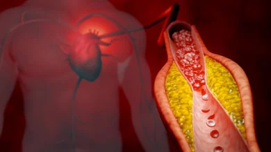
Congenital heart disease remains one of the most pervasive developmental challenges in global health today, impacting roughly two out of every one hundred newborns worldwide. Despite decades of intensive medical research, the precise molecular triggers responsible for the malformation of the heart during embryonic stages have largely eluded scientists. However, a multidisciplinary team of researchers at the University of Copenhagen has now shed significant light on this enduring medical mystery. By identifying a previously unknown cellular communication system operating on the exterior of embryonic cells, these scientists have opened a critical new frontier in understanding—and potentially one day treating—syndromic congenital heart conditions.
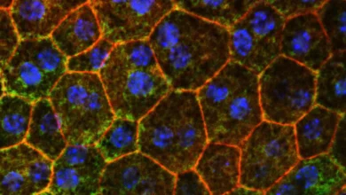
The breakthrough, published in the scientific journal PLOS Biology, centers on a microscopic, antenna-like structure known as the primary cilium. Projecting outward from the surface of nearly all human cells, this cellular antenna acts as an essential sensory organ, detecting chemical cues from the surrounding cellular microenvironment and translating those external messages into internal operational commands. These instructions dictate vital cell behaviors, including replication, directional migration, metabolic adjustment, and programmed cell death.
Within this microscopic antenna, the research team discovered a specialized signaling hub composed of three distinct proteins: TAK1, TAB2, and PKA-Cα. Working in tandem, these proteins serve as critical molecular switchboard operators, guiding stem cells on precisely when and how to differentiate into healthy heart muscle tissue. When genetic mutations disrupt this delicate signaling network, the resulting structural and functional defects in the cellular antenna can derail normal embryonic development, culminating in structural heart abnormalities.
The Path of Discovery: A Timeline and Multidisciplinary Approach
The journey toward this landmark discovery was neither short nor straightforward, reflecting years of rigorous, collaborative investigation across multiple scientific disciplines. The research initiative began with a comprehensive genetic screening of several thousand clinical patients diagnosed with congenital heart defects. By analyzing rare genetic variations and contrasting their frequency among patients versus healthy control populations, the researchers were able to pinpoint specific genetic anomalies strongly associated with the disease.
Following the human genetic analysis, the timeline shifted toward functional validation in the laboratory. To determine whether the identified genetic variants directly caused structural cardiac problems, the team deployed advanced genetic engineering techniques to replicate the human mutations in zebrafish models. Observing the embryonic development of these zebrafish revealed that alterations in the target genes severely compromised normal cardiac formation and measurably reduced overall heart function.
To dissect the cellular machinery with even greater precision, the scientists subsequently conducted intricate in vitro experiments utilizing human and mouse stem cell models. This multi-tiered methodological approach allowed the team to cross-verify their findings across diverse biological systems. Because the genetic associations observed in clinical patients aligned seamlessly with the experimental outcomes in zebrafish and mammalian stem cells, the investigators expressed a high degree of confidence that this newly mapped cellular mechanism operates identically in human embryonic development.
Anatomy of a Global Health Challenge
To fully appreciate the significance of the University of Copenhagen study, one must examine the broader epidemiological landscape of congenital heart disease. According to data compiled by the World Heart Federation and the Danish Heart Foundation, between 2.3 and 2.5 million newborns are diagnosed with structural heart abnormalities annually on a global scale. By 2023, epidemiological estimates indicated that approximately 16 million individuals were living with these lifelong conditions.
Clinically, these abnormalities are broadly categorized into two primary groups: syndromic and non-syndromic congenital heart disease. Non-syndromic cases occur as isolated structural heart defects without accompanying developmental anomalies elsewhere in the body. Conversely, syndromic congenital heart disease—the specific focus of the University of Copenhagen study—involves heart defects that manifest as part of a wider, more complex genetic syndrome affecting multiple organ systems simultaneously.
Historically, treating and counseling patients with syndromic congenital heart defects has posed immense challenges for pediatric cardiologists and geneticists, largely because clinicians struggled to find a single, unifying pathological explanation for why a child might present concurrently with heart malformations, neurological impairments, and skeletal or renal abnormalities.
Expert Insights and Mechanistic Implications
The implications of the newly discovered ciliary signaling hub extend far beyond pediatric cardiology, offering potential explanations for a wide array of congenital anomalies.
"We have discovered a new communication system on the exterior of the cell that is crucial for the proper formation of the heart during embryonic development," notes Lars Allan Larsen, an expert in congenital heart disease and Professor at the Department of Cellular and Molecular Medicine at the University of Copenhagen. "This finding changes our understanding of why some congenital heart defects arise. You could say that we have identified an important cog in a highly complex machine."
Larsen emphasizes the robustness of the team’s methodology, noting that the convergence of human genetic data, zebrafish models, and stem cell assays provides a compelling evidentiary foundation. "We investigate the mechanism from many different angles and using many different methods, all of which support what we observe in patients. Therefore, we are reasonably confident that this mechanism also exists in humans."
Sören Tvorup Christensen, Professor of cell biology at the Department of Biology and co-author of the study, elaborates on the precise molecular function of the proteins involved. "These proteins act as molecular instructions that tell stem cells when and how to develop into heart muscle cells. However, genetic alterations can disrupt this communication, causing ‘antenna defects’, which may lead to congenital heart defects."
Christensen also highlights the systemic nature of primary cilia dysfunction, noting that when this ciliary mechanism fails during embryogenesis, the disruption rarely remains isolated to a single tissue. "When the ciliary mechanism fails, it typically affects the development of several other organs as well. This may explain why some patients with congenital heart disease also have defects and related conditions affecting the brain, kidneys and skeleton. The mechanism provides a unifying explanation for diseases that we have previously struggled to understand."
Broadening Horizons: Beyond the Heart
Because primary cilia are distributed across nearly every cell type in the human body and play foundational roles in the morphogenesis of the brain, skeleton, and internal organs, uncovering the mechanics of the TAK1-TAB2-PKA-Cα signaling hub holds immense promise for the broader field of ciliopathies—genetic disorders linked to defective cilia.
Numerous rare genetic conditions are currently known to stem from ciliary dysfunction, yet the precise biochemical pathways driving disease progression have remained murky. By clarifying how minor genetic alterations within the primary cilium can trigger widespread multi-organ developmental failures, this research paves the way for advanced diagnostic frameworks.
Looking toward the future, the research team anticipates that this foundational knowledge will eventually streamline clinical protocols, enabling physicians to identify at-risk patients much earlier in life. Furthermore, by isolating the specific molecular signaling pathways responsible for cellular differentiation during embryogenesis, pharmaceutical researchers may one day be positioned to design targeted molecular therapies or preventative interventions for expectant mothers carrying genetic risk factors.
While immediate clinical applications will require further translation research and validation in human clinical trials, the study authored by Christensen, Larsen, and their colleagues marks a definitive milestone in molecular biology. By illuminating the microscopic antenna that guides the beating heart from its earliest embryonic origins, science has taken a monumental step toward decoding the fundamental blueprints of human life.
The complete research findings are available in the scientific journal PLOS Biology. The study was conducted by an extensive research team from the University of Copenhagen, including Sören Tvorup Christensen, Lars Allan Larsen, Canan Doganli, Oskar Kaaber Thomsen, Daniel A. Baird, Yeasmeen Ali, Menachem V. K. Sarusie, Line Jeanett Jessen, Pauline Munck Truelsen, Johanne Bay Mogensen, Maria Schröder Holm, Lorenzo Buttó, Maria Diamanti, Jindřich Leischner Fialová, and Lotte Bang Pedersen.