
Understanding Pelvic Organ Prolapse and the Clinical Implications of Cervical Protrusion in Women

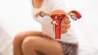
The sensation of a protruding cervix, often described by patients as a physical bulge or pressure within the vaginal canal, serves as a significant clinical indicator for various gynecological conditions, most notably Pelvic Organ Prolapse (POP). While the cervix—the lower, narrow portion of the uterus that connects to the vagina—naturally fluctuates in position and firmness throughout a woman’s menstrual cycle, a persistent protrusion beyond the vaginal opening or a significant descent within the canal requires comprehensive medical evaluation. According to the American College of Obstetricians and Gynecologists (ACOG), this condition occurs when the pelvic floor muscles and connective tissues, such as ligaments and fascia, become weakened or damaged, losing their ability to support the pelvic organs.
Pelvic organ prolapse is a multifaceted condition that can involve the uterus, bladder, or rectum. When the uterus descends, the cervix is pushed downward, leading to what is commonly referred to in layman’s terms as a "fallen womb." Medical professionals emphasize that while the condition is rarely life-threatening, its impact on a woman’s quality of life, including physical comfort, sexual health, and urinary function, can be profound. Understanding the etiology, stages of progression, and modern diagnostic standards is essential for early intervention and effective management.
The Pathophysiology and Mechanism of Pelvic Floor Descent
The pelvic floor is a complex arrangement of muscles and connective tissues that forms a "hammock" across the base of the pelvis. This structure supports the pelvic organs, including the uterus, vagina, bladder, and bowel. The integrity of this hammock is maintained by the levator ani muscle group and the endopelvic fascia. When these structures are compromised, the internal organs begin to shift due to the constant force of intra-abdominal pressure and gravity.
The chronology of prolapse usually begins with subtle stretching of the ligaments. Over time, particularly following repetitive strain or significant life events such as childbirth, these tissues may lose their elasticity. As the support fails, the uterus begins its descent. In the early stages, the cervix may only sit lower in the vagina than usual. In advanced stages, the cervix and even the entire body of the uterus can protrude through the introitus (the vaginal opening). This progression is often categorized into stages ranging from Stage I (slight descent) to Stage IV (total procidentia, where the organ is entirely outside the body).
Primary Causes and Risk Factors for Cervical Protrusion
Medical research identifies several key factors that contribute to the weakening of the pelvic support system. The most prevalent cause is pregnancy and vaginal childbirth. During labor, the pelvic floor muscles and nerves are subjected to extreme stretching and potential trauma. Large birth weights (macrosomia) and prolonged pushing phases are specifically linked to a higher risk of future prolapse.
However, childbirth is not the sole factor. Aging and the onset of menopause play a critical role. As women age, the natural production of estrogen—a hormone vital for maintaining the strength and thickness of pelvic connective tissues—decreases. This hormonal shift leads to the thinning of the vaginal walls and the weakening of the pelvic ligaments.
Beyond biological factors, chronic intra-abdominal pressure is a significant contributor. This includes:
- Obesity: Excess weight places constant pressure on the pelvic floor.
- Chronic Respiratory Issues: Persistent coughing, often seen in smokers or those with asthma, creates repetitive downward pressure.
- Chronic Constipation: Frequent straining during bowel movements can overstretch the pelvic nerves and muscles over time.
- Heavy Lifting: Occupations or fitness routines involving the repetitive lifting of heavy objects without proper core engagement can accelerate the descent of pelvic organs.
Diagnostic Standards and the POP-Q System
A definitive diagnosis of pelvic organ prolapse cannot be made through self-examination alone. While a woman may feel a lump or "heaviness," clinical verification is required to determine the severity and the specific organs involved. Gynecologists typically utilize the Pelvic Organ Prolapse Quantification (POP-Q) system, an international standard for measuring the degree of descent.
During a POP-Q examination, the physician takes several measurements of the vaginal canal and the position of the cervix relative to the hymen. The patient may be asked to perform a Valsalva maneuver (straining as if having a bowel movement) or cough while in a lithotomy or standing position. This allows the clinician to observe the maximum extent of the prolapse under pressure. This standardized approach ensures that treatment plans are based on objective data rather than subjective sensations.
Supporting Data and Global Prevalence
Epidemiological data highlights the scale of this health issue. Studies published in the International Urogynecology Journal suggest that nearly 50% of women who have given birth will experience some degree of pelvic organ prolapse in their lifetime, though many will remain asymptomatic. In the United States, approximately 3% to 6% of women report symptomatic prolapse, but this number is expected to rise as the global population ages.
In Indonesia and other developing nations, the prevalence may be influenced by different factors, including the number of pregnancies per woman and the availability of postpartum pelvic floor rehabilitation. Research indicates that early-stage prolapse often goes undiagnosed due to cultural stigmas surrounding gynecological health, leading many women to seek help only when the condition has reached Stage III or IV.
Clinical Manifestations and Patient Symptoms
The symptoms of a protruding cervix or prolapsed uterus are often described as a "dragging" sensation in the pelvis. Patients may feel as though they are sitting on a small ball. Other common clinical manifestations include:
- Urinary Incontinence: Involuntary leakage of urine during coughing, sneezing, or exercise.
- Incomplete Voiding: The feeling that the bladder or bowels have not fully emptied.
- Dyspareunia: Pain or discomfort during sexual intercourse.
- Vaginal Bleeding or Discharge: Occurs when the protruding tissue rubs against clothing, leading to irritation or ulceration of the cervical mucosa.
- Lower Back Pain: Often worsening as the day progresses due to the cumulative effects of gravity on the unsupported organs.
Treatment Modalities: From Conservative to Surgical
Management of a protruding cervix is highly individualized, depending on the severity of the prolapse, the patient’s age, her desire for future pregnancies, and the impact on her daily life.
1. Conservative Management:
For mild to moderate cases (Stage I and II), Pelvic Floor Muscle Training (PFMT), commonly known as Kegel exercises, is the first line of defense. When performed correctly and consistently, these exercises can strengthen the levator ani muscles and prevent further descent. Additionally, the use of a vaginal pessary—a removable silicone device inserted into the vagina to provide structural support—is a highly effective non-surgical option.
2. Surgical Intervention:
In advanced cases or when conservative measures fail, surgery may be necessary. Options include:
- Reconstructive Surgery: Aimed at repairing the weakened fascia and "re-suspending" the uterus using the patient’s own tissues or synthetic mesh.
- Obliterative Surgery: A procedure (colpocleisis) that narrows or closes the vagina to support the organs. This is typically reserved for elderly patients who are no longer sexually active.
- Hysterectomy: In some instances, removing the uterus is part of the repair process, although uterus-sparing techniques are increasingly favored in modern urogynecology.
Implications for Public Health and Quality of Life
The implications of untreated pelvic organ prolapse extend beyond physical discomfort. There is a significant psychological component; many women experience anxiety, body image issues, and a withdrawal from social or physical activities due to the fear of leakage or the discomfort of the protrusion.
Medical experts advocate for a shift in postpartum care, emphasizing the importance of pelvic floor physical therapy as a standard of recovery. By addressing minor muscle weaknesses early, the long-term risk of severe prolapse can be mitigated. Furthermore, public health initiatives aimed at reducing obesity and improving nutritional standards to prevent constipation can serve as primary prevention strategies for pelvic floor disorders.
Expert Recommendations and Conclusion
For women who notice a change in the position of their cervix or feel a protrusion, the clinical recommendation is clear: seek a professional evaluation. Early detection through a pelvic exam can differentiate between normal physiological changes, such as those occurring during the menstrual cycle or pregnancy, and the onset of pelvic organ prolapse.
The evolution of urogynecological medicine has provided women with a wide array of options to manage and correct cervical protrusion. From advanced physical therapy techniques to minimally invasive robotic surgeries, the goal remains the restoration of anatomy and function. As the medical community continues to refine the POP-Q system and surgical materials, the prognosis for women with pelvic organ prolapse continues to improve, allowing for a return to an active, symptom-free lifestyle. Diagnosis is the first step toward reclaiming pelvic health, and women are encouraged to discuss these symptoms openly with their healthcare providers to avoid the progression of a condition that, while common, should not be accepted as an inevitable part of aging.