The Sweet Signal: How Fructose Metabolism Drives Aggressive Ovarian Cancer Metastasis After Chemotherapy

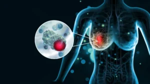
A groundbreaking study conducted at The Wistar Institute has unveiled a sophisticated, previously unknown mechanism by which aggressive ovarian cancer persists and spreads within the human body. Published in the journal Nature Aging, the research identifies a startling culprit in the post-chemotherapy survival strategy of tumor cells: fructose. This common sugar, often found in high concentrations in processed foods and sweetened beverages, appears to function as a chemical messenger that triggers neighboring cancer cells to detach from the primary tumor, facilitating metastasis throughout the abdominal cavity.
The Challenge of Ovarian Cancer Recurrence
Ovarian cancer remains one of the most lethal gynecological malignancies, primarily due to its tendency to recur even after aggressive medical intervention. Standard treatment protocols for the disease typically involve platinum-based chemotherapy. These drugs work by inducing DNA damage within rapidly dividing cancer cells, effectively preventing their proliferation.
While the majority of patients exhibit a positive initial response to these treatments, the clinical reality is sobering: most patients eventually experience a recurrence. When the cancer returns, it frequently manifests as widespread dissemination throughout the peritoneal cavity—the abdominal space housing vital organs like the liver, intestines, and stomach. This process, known as metastasis, is the primary driver of mortality in approximately 90% of ovarian cancer cases. For decades, the medical community has sought to understand why these seemingly dormant, chemotherapy-treated cells retain the capacity to incite secondary tumor growth.
The Biological Paradox: Active Yet Non-Dividing Cells
The research team at The Wistar Institute, led by Dr. Katherine Aird and postdoctoral fellow Dr. Aidan Cole, hypothesized that the "surviving" cancer cells were not merely passive bystanders after chemotherapy. Instead, they proposed that these cells remain biologically active in a senescence-like state, secreting a complex milieu of signaling molecules that alter the tumor microenvironment.
In a series of rigorous preclinical experiments, the researchers successfully isolated these surviving cells and analyzed their secretome—the collection of substances they release. The findings were definitive: even in the absence of the surviving cancer cells themselves, the molecules they secreted were sufficient to significantly escalate the spread of neighboring tumor cells. "As far as we know, this is the first time anyone has shown, in a preclinical model, that it’s the molecules these cells release—not the cells themselves—that drive the cancer’s spread," Dr. Cole noted.
Fructose as an Unexpected Signaling Molecule
The most surprising discovery during the molecular analysis was the role of fructose. Traditionally viewed by oncologists merely as an energy substrate for cellular metabolism, fructose was revealed to be a critical signal in the metastasis pathway. The study demonstrated that chemotherapy-stressed cells manufacture and export fructose, which then acts upon adjacent cancer cells to promote a "breakaway" phenotype.
The implications of this finding are profound when placed against the backdrop of modern dietary habits. In the United States, high-fructose corn syrup—a sweetener prevalent in carbonated beverages and processed snacks—constitutes a significant portion of the average caloric intake, sometimes ranging from 8% to 20% of daily calories. The researchers observed that high levels of fructose exposure, even in the absence of chemotherapy, could trigger signaling pathways associated with cancer cell migration. While the study does not suggest that sugar consumption directly causes the initial development of ovarian cancer, it raises urgent questions regarding the potential role of nutrition in modulating the progression and aggressiveness of established tumors.
The Mechanism: Cholesterol and Cellular Adhesion
To delineate the precise biological pathways involved, the team employed CRISPR-based gene editing technology to perform a systematic screen of cellular responses. They discovered that the fructose-induced signaling suppresses the synthesis of cholesterol within neighboring cancer cells.
While cholesterol is widely recognized for its association with cardiovascular health, its role in oncology is equally vital. Cholesterol is a structural cornerstone of cell membranes; it helps maintain the integrity of "cell-cell junctions"—the biological glues that keep cells anchored to one another. When fructose-mediated signaling inhibits cholesterol production, these junctions weaken. With their structural adhesion compromised, cancer cells find it significantly easier to detach, invade surrounding tissues, and migrate to distant sites within the abdominal cavity.
The Statin Connection: A Medical Nuance
The revelation that reduced cholesterol production facilitates cancer cell migration led the researchers to examine the potential side effects of statins, the world’s most commonly prescribed class of cholesterol-lowering medications. With approximately 39 million Americans currently using statins, the researchers hypothesized that these drugs might inadvertently mimic the effects of fructose-induced cholesterol suppression.
Their experimental data suggested that statins alone could indeed weaken the connections between cancer cells. This raises a complex clinical question: could cholesterol-lowering medication, when administered alongside chemotherapy, inadvertently create a more favorable environment for metastasis in some ovarian cancer patients?
"We haven’t tested this effect in patients yet, but it raises questions about combining cholesterol-lowering drugs with chemotherapy, especially since ovarian cancer is most common in postmenopausal women who are often already on statins," said Dr. Aird. The research team is careful to emphasize that these findings are preliminary and strictly experimental; patients currently prescribed statins for cardiovascular health should not discontinue their medication without consulting their primary care physicians.
Broader Implications and Future Research
While the primary focus of this study was ovarian cancer, the biological mechanisms identified may have far-reaching implications for other abdominal malignancies. The researchers suspect that pancreatic, colon, and liver cancers—all of which frequently spread throughout the torso—may share similar metabolic vulnerabilities.
The scientific community has responded to these findings with a mix of cautious optimism and a call for further clinical validation. Dr. Aird’s laboratory is already designing follow-up studies to determine if these metabolic pathways are universal across different tumor types. Furthermore, the potential to integrate nutritional strategies—such as diet modification—into the standard of care for cancer patients represents a frontier in personalized oncology.
However, experts stress that it is premature to issue broad dietary mandates based on this research. Establishing a causal link between dietary sugar intake and clinical outcomes in human patients will require long-term, multi-center longitudinal studies. Nevertheless, the identification of fructose as a signaling agent provides a novel therapeutic target. Future research may focus on developing pharmacological interventions that block the specific fructose-sensing receptors or the downstream pathways that lead to weakened cellular adhesion.
By shifting the focus from simply killing cancer cells to understanding the complex chemical conversations that occur within the tumor microenvironment, the Wistar Institute study provides a blueprint for a new era of cancer research. As investigators continue to unravel the roles of nutrients like fructose in disease progression, the medical community moves closer to a more nuanced understanding of how to prevent the recurrence of one of the most aggressive diseases in modern medicine. The findings serve as a reminder that the tumor microenvironment is a dynamic, nutrient-sensitive landscape, and that the intersection of diet, medication, and molecular biology may hold the key to stopping cancer in its tracks.