Mini Brain Organoids Offer Hope for Personalized Alzheimer’s Treatment and Diagnosis

Scientists at Johns Hopkins Medicine have unveiled groundbreaking research utilizing patient-derived brain organoids that could revolutionize how Alzheimer’s disease is treated and diagnosed. This innovative approach uses miniature, lab-grown models of brain tissue to predict individual patient responses to medications for neuropsychiatric symptoms, a common and often debilitating aspect of the disease. The findings, published in the esteemed journal Alzheimer’s & Dementia: The Journal of the Alzheimer’s Association, also suggest that tiny particles released by these organoids may serve as novel biomarkers for early diagnosis and disease progression.
Alzheimer’s disease, the most prevalent form of dementia, currently affects an estimated 7 million Americans, with global figures projected to rise significantly in the coming decades due to an aging population. The progressive neurodegenerative disorder is characterized by memory loss, cognitive decline, and behavioral changes. While there is no cure, managing its associated symptoms, particularly neuropsychiatric ones like anxiety, depression, agitation, and psychosis, is crucial for improving patients’ quality of life. However, the efficacy of current treatments, such as selective serotonin reuptake inhibitors (SSRIs), varies considerably among individuals, presenting a significant challenge for clinicians.
A Novel Approach: Patient-Derived Brain Organoids
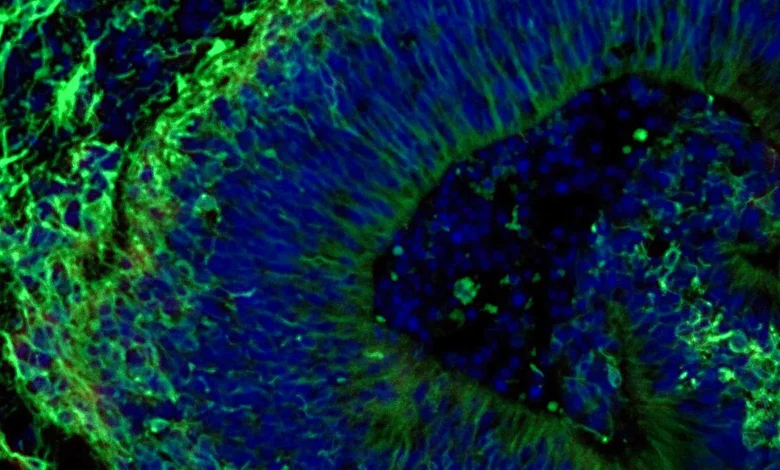
The core of this pioneering research lies in the creation of brain organoids—three-dimensional clusters of brain cells grown in a lab from a patient’s own cells. These "mini-brains" are designed to mimic specific aspects of the human brain, allowing researchers to study disease mechanisms and drug responses in a controlled environment. For this study, the Johns Hopkins team focused on hindbrain organoids, a region vital for regulating fundamental life functions such as breathing, sleep, and heart rate.
The process begins with a simple blood sample, ethically collected with patient consent from individuals diagnosed with Alzheimer’s disease, as well as from healthy control subjects. These blood cells are then reprogrammed into induced pluripotent stem cells (iPSCs). iPSCs possess the remarkable ability to differentiate into any cell type in the body, including specialized brain cells known as neurons. By coaxing these iPSCs to develop into neurons that produce serotonin, a key neurotransmitter involved in mood regulation, the researchers were able to construct pea-sized organoids that closely resemble segments of the human hindbrain. This study represents one of the largest-scale investigations of patient-derived brain organoids in Alzheimer’s research to date, incorporating hundreds of organoids representing a diverse range of patients and healthy individuals.
"Our study suggests that large-scale, patient-derived brain organoids and the vesicles they secrete can help us stage Alzheimer’s disease, investigate the mechanisms that drive it and assess how patient subgroups may respond to different treatments," stated Dr. Vasiliki Machairaki, the study’s lead investigator and an associate professor of genetic medicine at Johns Hopkins University School of Medicine. "There is currently no cure for Alzheimer’s disease. However, selective serotonin reuptake inhibitors (SSRIs) are often prescribed to help manage neuropsychiatric symptoms such as anxiety, depression, and agitation. These symptoms affect nearly all patients, but responses to the medications vary widely."
Uncovering Molecular Signatures of Alzheimer’s
Upon examination, the patient-derived organoids exhibited distinct molecular characteristics that mirrored key biological features of Alzheimer’s disease. Compared to organoids grown from the cells of healthy individuals, those derived from people with Alzheimer’s displayed significant differences in proteins crucial for neuronal communication, inflammatory responses, and pathways known to be implicated in the disease’s progression.
The researchers then introduced escitalopram oxalate, a widely prescribed SSRI, to these organoids. The results were illuminating. In some Alzheimer’s organoids, the medication triggered an increase in proteins associated with serotonin signaling and enhanced communication between brain cells—precisely the mechanisms targeted by antidepressants. However, in other organoids, the molecular response was minimal or entirely absent. This variability in response underscores the heterogeneity of Alzheimer’s disease and its impact on individual patients.
"We used these organoids to model how some patients’ tissue may respond to a commonly prescribed SSRI," Dr. Machairaki explained. "On a large-scale level, our model may eventually be used to identify subgroups of patients, based on underlying molecular mechanisms, who are more likely to respond to certain drugs and thus help us to create precise, targeted treatments in the long run." This breakthrough has profound implications for personalized medicine, moving away from a one-size-fits-all approach to treatment and towards therapies tailored to an individual’s unique biological profile.
Extracellular Vesicles: Potential Biomarkers for Diagnosis and Prognosis
Beyond observing drug responses in the organoids themselves, the research team delved into the role of extracellular vesicles (EVs). These are minuscule particles released by cells, carrying vital cellular information, including proteins and genetic material. The study found that EVs secreted by the brain organoids could potentially serve as valuable biomarkers for Alzheimer’s disease.
By analyzing the protein content of EVs released by both Alzheimer’s and healthy control organoids, both before and after escitalopram treatment, the scientists identified significant differences. The EVs from Alzheimer’s organoids contained altered levels of proteins critical for neuronal signaling, memory formation, and neurotransmitter release. Specifically, proteins like RAB3A, NSF, and ATCAY, essential for normal brain cell communication, were found at lower concentrations in EVs from Alzheimer’s organoids.
Crucially, the study observed that escitalopram treatment led to changes in the protein composition of these EVs, particularly in those linked to serotonin signaling and synaptic pathways. The extent of these changes mirrored the molecular response observed within the organoids, suggesting that EVs could offer a non-invasive window into how brain tissue is reacting to medication.
"This variation raises the possibility that extracellular vesicles from brain organoids could eventually help identify which patients are most likely to benefit from a particular treatment," Dr. Machairaki noted. The concept of using EVs as biomarkers is particularly exciting, as it opens the door to developing "liquid biopsies"—simple blood tests that could potentially diagnose Alzheimer’s disease, determine its severity, and even pinpoint specific disease subtypes. This would represent a monumental leap forward in early detection and personalized therapeutic strategies.
The Path Forward: Enhancing Organoid Realism and Clinical Application
The Johns Hopkins team is not resting on their laurels. Dr. Machairaki envisions developing even more sophisticated organoids that incorporate other crucial brain cell types, such as immune cells (microglia), and vascular networks that mimic blood vessels. The inclusion of these components would further enhance the organoids’ resemblance to living human brain tissue, leading to more accurate and comprehensive research findings.
The timeline for these advancements is still unfolding, but the implications are clear. With continued research and validation, EVs derived from brain organoids could one day revolutionize how Alzheimer’s disease is managed. The ability to predict drug efficacy before administering medication, coupled with the potential for early and precise diagnosis, could fundamentally alter the landscape of Alzheimer’s care, offering renewed hope to millions of patients and their families.
The study received significant support from the National Institutes of Health (NIH) and the Paul G. Allen Frontiers Foundation, underscoring the national and international importance placed on finding effective solutions for Alzheimer’s disease. The collaborative effort involved a multidisciplinary team of researchers from Johns Hopkins, the University of Rochester School of Medicine and Dentistry, and Tymora Analytical Operations.
Broader Impact and Implications for Alzheimer’s Research
The implications of this research extend far beyond the immediate scope of SSRI efficacy. The development of robust, patient-specific brain organoid models provides an unprecedented platform for exploring the complex, multifactorial nature of Alzheimer’s disease. Researchers can now investigate the intricate molecular pathways that lead to neuronal dysfunction and death, identify novel therapeutic targets, and screen potential drug candidates with a higher degree of precision.
The heterogeneity observed in the organoid responses highlights a critical aspect of Alzheimer’s: it is not a monolithic disease. Different individuals may have distinct underlying pathological mechanisms contributing to their cognitive decline and neuropsychiatric symptoms. By dissecting these variations at a molecular level, this research paves the way for precision medicine strategies tailored to specific Alzheimer’s subtypes.
Furthermore, the potential of extracellular vesicles as biomarkers could transform diagnostic paradigms. Current diagnostic methods often rely on cognitive assessments, imaging techniques like PET scans, and cerebrospinal fluid analysis, which can be invasive, costly, and may only detect the disease in its later stages. A simple blood-based liquid biopsy using EVs could enable earlier diagnosis, allowing for timely interventions and potentially slowing disease progression. It could also facilitate more accurate prognostication, helping clinicians and patients make informed decisions about care and future planning.
The timeline for widespread clinical application of these findings is still a matter of ongoing research and development. However, this study represents a significant stride forward, demonstrating the immense potential of advanced cellular models and biomarker discovery in the fight against Alzheimer’s disease. The Johns Hopkins team’s work not only advances our scientific understanding but also offers a tangible vision of a future where Alzheimer’s treatment and diagnosis are more precise, personalized, and ultimately, more effective.