
Early Adulthood Alcohol Use for Stress Relief Linked to Permanent Brain Alterations and Cognitive Decline in Middle Age
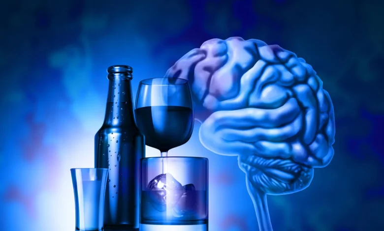
New research from the University of Massachusetts Amherst has uncovered evidence suggesting that the habit of using alcohol to manage stress during early adulthood may leave an indelible mark on the brain, persisting long after an individual has achieved sobriety. The study, which highlights a critical window of neurobiological vulnerability, indicates that these physiological changes often remain dormant until middle age, at which point they manifest as reduced mental flexibility and an increased susceptibility to relapse during periods of high tension. Perhaps most significantly, the research links these alcohol-induced brain modifications to the early stages of cognitive decline, potentially accelerating the onset of neurodegenerative conditions such as dementia and Alzheimer’s disease.
The findings, published in the peer-reviewed journal Alcohol: Clinical and Experimental Research, provide a sophisticated look at the intersection of substance use and psychological stress. By examining how these two factors interact to reshape neural circuits, the UMass Amherst team has challenged the traditional view that the brain can fully "reset" after long-term abstinence. Instead, the study suggests that the combination of heavy drinking and chronic stress during the formative years of early adulthood—typically the late teens through the twenties—reprograms the brain’s decision-making centers in ways that are difficult to reverse through behavioral changes alone.
The Neurobiological Mechanism of the Stress-Alcohol Cycle
For decades, clinicians and neuroscientists have observed a reciprocal relationship between alcohol consumption and stress. This "vicious cycle" is well-documented: individuals often turn to alcohol for its sedative properties to alleviate the symptoms of anxiety or environmental pressure. While alcohol may provide temporary relief by depressing the central nervous system, repeated exposure begins to erode the brain’s endogenous stress-management systems. Over time, the brain’s baseline for stress tolerance is lowered, requiring more alcohol to achieve the same level of calm—a phenomenon known as neuroadaptation.
However, the UMass Amherst study goes deeper into the specific neurocircuitry involved in this process. The research focused on the locus coeruleus (LC), a small but vital nucleus located in the brainstem. The LC is the brain’s primary source of norepinephrine, a neurotransmitter and hormone that mediates the body’s "fight or flight" response. In a healthy brain, the LC becomes highly active during a stressful event, facilitating rapid decision-making and heightened awareness, and then returns to a state of equilibrium once the threat has passed.
In the subjects studied—specifically mouse models chosen for their neurological similarities to humans—the researchers found that the dual impact of alcohol and chronic stress caused the LC to lose its "off switch." The molecular machinery required for the LC to regulate its own activity was significantly damaged. Consequently, the region remained in a state of perpetual dysregulation, impairing the animal’s ability to navigate complex or changing environments.
The Significance of Early Adulthood Exposure
The timing of alcohol exposure appears to be a critical factor in the severity of long-term damage. Early adulthood is a period of significant synaptic pruning and frontal lobe development in the human brain. When heavy alcohol use is introduced during this phase, particularly as a coping mechanism for stress, it interferes with the stabilization of neural pathways responsible for executive function.
The UMass Amherst study utilized a longitudinal approach to track these changes. By observing mice from their "early adult" phase into "middle age," the researchers were able to simulate the human experience of several decades. The data revealed that mice subjected to both stress and alcohol in their youth were significantly more likely to return to alcohol consumption when faced with new stressors in middle age, even if they had been "sober" for the majority of their lives.
"Middle age is when problems start to add up," noted Elena Vazey, an associate professor of biology at UMass Amherst and the study’s senior author. Her team’s findings suggest that the brain "remembers" the early-life association between alcohol and stress relief, creating a latent vulnerability that can be triggered by the natural challenges of aging.
Cognitive Flexibility vs. Learning Ability
One of the most striking findings of the research was the distinction between different types of cognitive performance. The study found that middle-aged mice with a history of stress-induced drinking showed no significant deficits in their basic learning ability. They were still capable of acquiring new information and performing routine tasks.
However, a profound deficit was observed in "cognitive flexibility." This refers to the brain’s ability to adapt to new rules or change strategies when a previous approach is no longer effective. In real-world human terms, cognitive flexibility is what allows a person to navigate a detour on a familiar route, adapt to a new software update at work, or manage complex interpersonal dynamics.
The loss of this flexibility is a hallmark of early-stage dementia. The inability to pivot or adjust to changing circumstances is often one of the first signs that the brain’s executive centers are failing. The fact that early adulthood drinking specifically targets this function suggests that it may be a primary driver of premature cognitive aging.
Oxidative Stress and the Link to Alzheimer’s Disease
The researchers also investigated the cellular health of the locus coeruleus and found high levels of oxidative stress. Oxidative stress occurs when there is an imbalance between the production of free radicals and the body’s ability to neutralize them with antioxidants. This imbalance leads to cellular damage, inflammation, and eventually, cell death.
Oxidative damage in the LC is a known precursor to Alzheimer’s disease. In humans, the LC is often the first brain region to show signs of protein aggregation—such as tau tangles—that are characteristic of Alzheimer’s. The UMass Amherst study found that even after long periods of abstinence, the middle-aged brains of formerly heavy-drinking mice showed little to no signs of repairing this oxidative damage.
This persistence suggests that the "scars" left by alcohol and stress are not merely functional but structural. The cells themselves are compromised, which explains why the brain struggles to recover its original level of performance. This finding provides a biological explanation for why some individuals experience cognitive decline earlier than others, even if they have maintained a healthy lifestyle in their later years.
Shifting the Paradigm of Addiction Treatment
The implications of this research for the treatment of Alcohol Use Disorder (AUD) are profound. Currently, many treatment programs focus heavily on the behavioral aspects of quitting—relying on willpower, therapy, and lifestyle changes to maintain sobriety. While these are essential components, the UMass Amherst study suggests they may be insufficient for those with a history of early-life stress-drinking.
"The brain’s wiring system is damaged, which means quitting drinking or making better decisions isn’t a matter of willpower," Vazey explained. If the brain’s hardware—the LC and its associated circuits—is physically altered, then the individual is operating with a "differently wired" system that may require medical or pharmacological intervention to repair.
This research supports the development of new therapeutic strategies that target oxidative stress and neuroinflammation directly. Future treatments might focus on neuro-restorative drugs that help the LC regain its regulatory function or antioxidants that can cross the blood-brain barrier to mitigate the damage caused by early-life drinking.
Supporting Data and Global Context
The UMass Amherst study aligns with broader public health data regarding alcohol and aging. According to the National Institute on Alcohol Abuse and Alcoholism (NIAAA), which supported the study, alcohol-related brain damage is a leading cause of preventable cognitive impairment. Statistics show that individuals with a history of AUD are twice as likely to develop some form of dementia compared to those who drink in moderation.
Furthermore, the World Health Organization (WHO) has identified a rising trend in "binge drinking" and high-stress levels among young adults globally. As this cohort moves into middle age, public health systems may face a significant increase in early-onset cognitive disorders. The timeline suggested by the UMass Amherst research indicates that the "bill" for early adulthood substance use often comes due 20 to 30 years later, making early intervention and education vital.
Conclusion and Future Directions
The study from the University of Massachusetts Amherst provides a sobering look at the long-term consequences of using alcohol as a crutch for stress. By identifying the locus coeruleus as a primary site of damage and highlighting the persistence of oxidative stress, the research offers a new roadmap for understanding the link between addiction and neurodegeneration.
As the scientific community continues to explore these connections, the focus is likely to shift toward early screening and the identification of biomarkers for brain health. Understanding that the brain "remembers" the stresses and substances of our youth serves as a powerful reminder of the importance of developing healthy coping mechanisms early in life. For those who have already moved past their drinking years, the research underscores the need for a medical approach to sobriety—one that views brain health as a lifelong project of restoration rather than just a test of character.


