In a significant breakthrough for regenerative medicine and orthopedics, a collaborative team of researchers from Weill Cornell Medicine and the Hospital for Special Surgery (HSS) has successfully isolated a previously elusive population of stem cells responsible for the formation and maintenance of tendons and ligaments. This discovery, published in the September 7 issue of the journal Cell, provides a long-sought explanation for how the body sustains the connective tissues that bridge the gap between muscle and bone. More importantly, the research suggests that these specialized cells may play a pivotal, pathological role in the development of lumbar spinal stenosis, a debilitating condition that affects an estimated 103 million individuals globally.
The identification of these cells—which function as the master builders for tendons and ligaments throughout the human body—marks the culmination of years of investigative work into skeletal stem cell biology. By isolating the cellular origin of these tissues, the research team has opened a new frontier for non-surgical interventions, potentially shifting the clinical paradigm for millions of patients currently reliant on invasive procedures to address spinal nerve compression.
A History of Skeletal Discovery
The path to this discovery was paved by a series of foundational breakthroughs led by Dr. Matthew Greenblatt, a co-corresponding author of the study and associate professor of pathology and laboratory medicine at Weill Cornell. Dr. Greenblatt’s laboratory has long focused on mapping the diverse landscape of skeletal stem cells.
The research chronology began in earnest in 2018, when the team successfully identified the stem cell population responsible for initiating fracture repair within the outer layer of bone. Following this success, the researchers systematically widened their search, discovering distinct stem cell populations involved in the formation of the skull and the spine. However, identifying the equivalent stem cells for tendons and ligaments presented a unique set of technical hurdles. Unlike bone, which possesses a more distinct cellular architecture, tendons and ligaments are densely packed with fibroblast-like cells. These cells are notoriously difficult to differentiate under standard microscopic analysis, as their morphological profiles are remarkably similar, effectively masking the existence of a rare, underlying stem cell population.
To overcome this, Dr. Greenblatt and his team, including first author Dr. Lingling Hu, employed advanced single-cell analysis techniques. By analyzing thousands of individual cells and sorting them into distinct populations, the researchers were able to isolate the specific subset that exhibited "stemness"—the capacity to self-renew while simultaneously generating the full spectrum of mature cells required for tendon and ligament tissue homeostasis.
The Mechanism of Spinal Stenosis
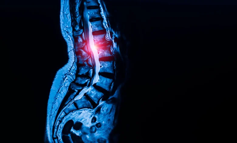
The clinical relevance of this discovery became apparent when the team examined the role of these stem cells in the context of lumbar spinal stenosis. This condition occurs when the spinal canal narrows, typically due to the thickening of ligaments, which exerts painful pressure on the nerves. Symptoms frequently include chronic back pain, numbness, and severe mobility impairment, often requiring surgery to decompress the spinal cord when conservative treatments fail.
To determine if the newly identified stem cells were complicit in this pathology, the researchers conducted a comparative analysis. They collected ligament tissue from two groups of patients undergoing surgery: those suffering from severe spinal stenosis and a control group suffering from herniated discs without signs of stenosis.
The results were striking. Ligaments harvested from patients with stenosis contained a significantly higher density of these stem cells compared to the control group. When these "stenotic" stem cells were transplanted into mice, they exhibited an aggressive tendency to produce excess tendon tissue, confirming that the cells were not merely present in higher numbers but were actively driving the structural thickening that characterizes the disease.
Further investigation into the internal signaling pathways of these cells revealed that they are governed by calcium signaling. In the stenotic cells, researchers observed hyperactive calcium signaling, which acts as a molecular "gas pedal" for cell growth and tissue formation. When the team experimentally increased this signaling in healthy cells, it triggered the same overgrowth patterns observed in the disease state. Conversely, by inhibiting these pathways in a mouse model, the researchers successfully blocked the abnormal tissue expansion.
Clinical Implications and Therapeutic Potential
The discovery that calcium signaling regulates the activity of these stem cells offers a tantalizing prospect for pharmacology. Because calcium channel blockers are already well-established medications used to treat hypertension, they represent a class of drugs with known safety profiles that could potentially be repurposed for the treatment of spinal stenosis.
"Identifying these specialized stem cells unlocks a new area of research that allows us to address this disease much more mechanistically, rather than just waiting until a patient’s condition worsens and requires surgery to relieve the nerve compression," stated Dr. Sravisht Iyer, co-corresponding author and a spine surgeon at HSS. "The findings are exciting for their potential to change the way we deliver spinal care."
While the prospect of a pharmaceutical treatment is highly encouraging, the researchers emphasize that clinical trials are essential to determine the efficacy and safety of repurposing these drugs for orthopedic use. If successful, such a treatment could offer a non-invasive alternative for the millions of people who currently face limited options once their spinal condition advances.
Broader Biological Significance
The reach of this discovery extends far beyond the spine. During their investigation, the researchers confirmed the presence of these stem cells in diverse connective tissues, including the kneecap ligament and the Achilles tendon. This suggests that the identified cell population is, in fact, the "universal" stem cell for all tendons and ligaments throughout the human body.
This universality creates a ripple effect across several fields of medicine. Future research initiatives are already being conceptualized to explore the role of these cells in other connective tissue disorders, such as Marfan syndrome, a genetic condition that impacts the structural integrity of tissues throughout the body. Additionally, the discovery may provide long-awaited insights into the underlying causes of chronic sports injuries, such as rotator cuff tears and persistent tendon degeneration, which have historically been difficult to treat due to poor natural healing capacity.
"Given that this cell appears to be the ultimate origin of all tendon and ligament cells, defects in this cell are likely at the heart of a wide range of tendon and ligament disorders," Dr. Greenblatt noted.
Conclusion and Future Outlook
The research, which was supported by a wide array of prestigious organizations including the National Institutes of Health, the Marfan Foundation, and the Arthritis National Research Foundation, represents a foundational shift in how medical science views connective tissue health. By moving from a symptomatic, surgical-centric model to one that targets the cellular origins of tissue growth, the medical community may be on the cusp of preventing the debilitating progression of common orthopedic diseases.
As the scientific community begins to digest these findings, the next phase of research will likely focus on the precise genetic and environmental triggers that cause these universal stem cells to become overactive. For the millions of patients living with the daily pain of spinal stenosis, the prospect of an early-intervention, medication-based treatment represents a profound evolution in the standard of care. With this discovery, Weill Cornell and HSS have not only identified the building blocks of the human frame but have also provided a blueprint for how to repair them when they go awry.
Related posts: