
NTU Singapore Scientists Identify Brain Waste Drainage Blockages as Crucial Early Warning Sign for Alzheimer’s Disease in Asian Populations
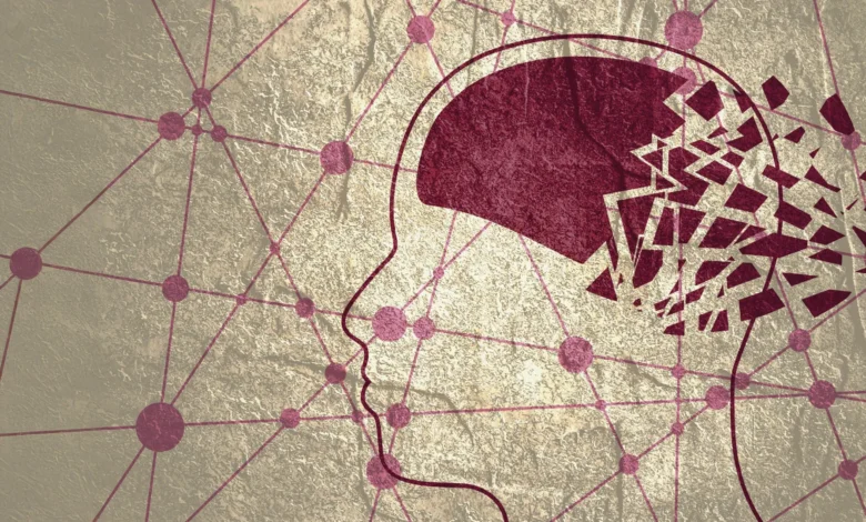
Scientists from Nanyang Technological University, Singapore (NTU Singapore) have uncovered a significant neurological marker that could revolutionize the early detection of Alzheimer’s disease. The research, led by the Lee Kong Chian School of Medicine (LKCMedicine), reveals that the brain’s waste removal system frequently becomes obstructed in individuals exhibiting early signs of cognitive decline. These blockages, visible on standard medical imaging, appear well before the onset of definitive dementia symptoms, offering a vital window for medical intervention.
The study focuses on "enlarged perivascular spaces" (EPVS), which act as the brain’s primary drainage channels. When these pathways become clogged, the brain loses its ability to clear toxic metabolic byproducts, leading to the accumulation of harmful proteins associated with neurodegeneration. By identifying these anomalies through routine Magnetic Resonance Imaging (MRI) scans, clinicians may soon be able to diagnose Alzheimer’s risk earlier and more affordably than current gold-standard methods allow.
The Biological Mechanism of Brain Waste Clearance
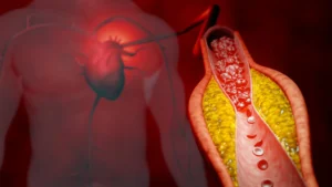
To understand the significance of the NTU discovery, it is essential to examine the glymphatic system—the brain’s unique waste-clearance pathway. Unlike the rest of the body, which relies on the lymphatic system to remove cellular debris, the brain utilizes perivascular spaces. These are fluid-filled gaps surrounding the blood vessels that penetrate the brain tissue. They serve as a plumbing system, flushing out metabolic waste products including beta-amyloid and tau proteins.
In a healthy brain, these proteins are efficiently drained away. However, in patients predisposed to Alzheimer’s, these channels can become "clogged" or enlarged. As the drainage efficiency drops, the perivascular spaces dilate, becoming large enough to be detected as small spots on an MRI scan. Until this study, the scientific community remained uncertain whether these enlarged spaces were a mere byproduct of aging or a direct precursor to Alzheimer’s disease. The NTU team’s findings strongly suggest the latter, positioning EPVS as a proactive diagnostic tool rather than a reactive observation.
Addressing the Data Gap in Asian Populations
One of the most critical aspects of this research is its focus on Asian demographics. Historically, the vast majority of Alzheimer’s research has been conducted on Caucasian populations in North America and Europe. This geographic bias has created a significant gap in global medical knowledge, as neurodegenerative diseases do not manifest identically across different ethnic groups.
Associate Professor Nagaendran Kandiah, the study’s lead and Director of the Dementia Research Centre (Singapore) at LKCMedicine, highlighted the genetic disparities that necessitate region-specific research. For instance, the apolipoprotein E4 (APOE4) gene is a well-known risk factor for Alzheimer’s. In Caucasian populations, approximately 50 to 60 percent of dementia patients carry this gene. Conversely, in Singapore, less than 20 percent of dementia patients are APOE4 carriers.
These statistics underscore the fact that the underlying drivers of Alzheimer’s can vary significantly by population. By studying nearly 1,000 Singaporean participants from diverse ethnic backgrounds—including Chinese, Malay, and Indian—the NTU team has provided a more accurate blueprint for diagnosing the disease in Asian contexts. This localized data is essential for developing precision medicine strategies that are effective for the world’s most populous continent.
Methodology: A Multi-Modal Approach to Cognitive Health
The study involved a comprehensive analysis of 1,000 participants, categorized into those with normal cognitive function and those experiencing Mild Cognitive Impairment (MCI). MCI is often considered the "twilight zone" of cognitive health—a stage where individuals experience noticeable memory or thinking difficulties that are not yet severe enough to interfere with daily independence, but which frequently progress to full-blown dementia.
The researchers utilized two primary diagnostic pillars:
- Neuroimaging: High-resolution MRI scans were used to visually identify and quantify enlarged perivascular spaces and white matter hyperintensities (damage to the brain’s "wiring").
- Blood Biomarkers: The team measured seven specific biochemicals in the blood, including various forms of beta-amyloid and tau proteins. These blood-based markers are currently at the cutting edge of Alzheimer’s research, offering a less invasive alternative to lumbar punctures (spinal taps) or expensive PET scans.
By correlating the physical blockages seen on MRIs with the chemical "red flags" in the blood, the researchers were able to establish a direct link between clogged brain drains and the molecular hallmarks of Alzheimer’s.
Key Findings: EPVS as a Superior Early Warning Signal
The results of the analysis were definitive. Participants diagnosed with Mild Cognitive Impairment were significantly more likely to exhibit enlarged perivascular spaces compared to their cognitively healthy counterparts. Furthermore, the presence of these enlarged spaces was strongly associated with four out of the seven Alzheimer’s-related blood markers.
A particularly striking discovery emerged when comparing EPVS to white matter damage. Traditionally, white matter hyperintensities—visible as "white spots" on an MRI—have been the standard marker used by doctors to assess vascular damage and dementia risk. While white matter damage did correlate with six of the seven blood markers, the NTU team found that in patients with early cognitive decline, the link between biochemical Alzheimer’s markers and enlarged perivascular spaces was actually stronger.
This suggests that EPVS may be a more sensitive and earlier indicator of Alzheimer’s pathology than the markers currently used in clinical practice. Because these spaces can be identified on routine MRI scans performed for general cognitive complaints, they provide a "free" layer of diagnostic data that does not require additional specialized testing or high costs for the patient.
Perspectives from the Medical Community
The implications of the study have drawn praise from across Singapore’s medical landscape. Justin Ong, a fifth-year LKCMedicine student and the study’s first author, emphasized the human element of the discovery. He noted that every month or year gained through early detection represents more time for patients and families to plan, and more time for medical interventions to slow the erosion of memory and personality.
Dr. Rachel Cheong Chin Yee, a Senior Consultant at Khoo Teck Puat Hospital’s Department of Geriatric Medicine, remarked on the significance of small blood vessel changes. She noted that identifying high-risk individuals before symptoms appear is the "holy grail" of dementia care, and this study moves the needle closer to that goal.
Dr. Chong Yao Feng, a Consultant at the National University Hospital’s Division of Neurology, pointed out that the study challenges the traditional separation of cerebrovascular disease (problems with blood vessels) and Alzheimer’s disease (a neurodegenerative condition). The research demonstrates a synergistic relationship: when the "pipes" (perivascular spaces) fail, the "waste" (amyloid and tau) builds up, accelerating the disease. This holistic view of brain health suggests that treating vascular health could be just as important as targeting protein plaques in the fight against Alzheimer’s.
Clinical Implications and Economic Impact
From a clinical standpoint, the ability to use routine MRIs for Alzheimer’s screening is a game-changer. Currently, confirming an Alzheimer’s diagnosis often requires a Positron Emission Tomography (PET) scan, which can cost thousands of dollars and requires the injection of radioactive tracers, or a lumbar puncture, which is invasive and often avoided by patients.
If enlarged perivascular spaces become a standard diagnostic metric, the cost of screening could drop precipitously. This is particularly important in the context of an aging global population. By 2050, the number of people living with dementia worldwide is expected to triple. Early detection allows for the implementation of lifestyle changes—such as diet, exercise, and cognitive training—and the use of emerging pharmaceutical treatments that are most effective in the early stages of the disease.
Timeline of Research and Future Directions
The NTU study represents a significant milestone in a multi-year effort to understand dementia in Singapore. The research was conducted as part of the Scholarly Project module in LKCMedicine’s Bachelor of Medicine and Bachelor of Surgery programme, demonstrating the high level of clinical research being integrated into medical education in Singapore.
The next phase of the research involves a longitudinal study. The NTU team plans to follow the same group of participants over several years to track the progression of their cognitive health. This will allow the scientists to confirm exactly how many individuals with enlarged perivascular spaces eventually develop Alzheimer’s dementia.
"Our findings carry substantial clinical implications," said Assoc Prof Kandiah. "While white matter damage is more widely used today because it is easily recognized, our results suggest that enlarged perivascular spaces hold unique value. They are not just markers of age; they are markers of a system in failure."
Conclusion: A New Frontier in Neurodegenerative Diagnostics
The work of the NTU Singapore team provides a powerful new lens through which to view Alzheimer’s disease. By shifting the focus toward the brain’s waste removal infrastructure, they have identified a physical precursor to the chemical changes that define the disease.
For the medical community, particularly in Asia, this research provides a culturally and genetically relevant framework for tackling a growing health crisis. As diagnostic technology evolves, the integration of MRI-based EPVS assessment could become a routine part of geriatric care, ensuring that the "clogged drains" of the brain are identified and addressed long before the lights of memory begin to fade. This study does more than just identify a biomarker; it provides a roadmap for earlier intervention, potentially changing the trajectory of life for millions of aging individuals.


