Cellular Antennas Hold the Key to Unlocking the Mysteries of Congenital Heart Disease, University of Copenhagen Researchers Discover


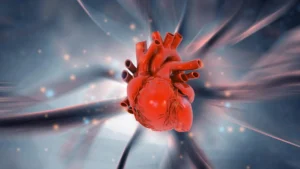
Congenital heart disease remains one of the most persistent medical challenges in modern pediatrics and developmental biology. Affecting approximately two out of every 100 newborns worldwide, these structural abnormalities of the heart develop during the earliest stages of embryonic growth. Despite decades of intense medical research, the exact molecular triggers that cause these defects to form have largely remained elusive. However, a major breakthrough by a multidisciplinary team of researchers at the University of Copenhagen has shed new light on this pervasive health issue, identifying a previously unknown cellular communication mechanism that governs proper heart formation.
The study, recently published in the scientific journal PLOS Biology, pinpoints a microscopic, antenna-like structure protruding from the surface of cells—known as the primary cilium—as a critical signaling hub. By demonstrating how specific proteins within this cellular antenna coordinate the transformation of stem cells into healthy heart muscle tissue, the researchers have uncovered a foundational cog in the exceptionally complex machinery of human development. This discovery not only reshapes scientific understanding of congenital heart defects but also opens new avenues for diagnosing and potentially treating a wide array of rare genetic disorders that affect multiple organ systems.
The Scale of the Crisis: Understanding Congenital Heart Defects
To fully appreciate the significance of the University of Copenhagen discovery, it is essential to examine the profound global impact of congenital heart disease. According to data from the World Heart Federation and the Danish Heart Foundation, between 2.3 and 2.5 million newborns are diagnosed with congenital heart defects each year. Globally, an estimated 16 million people were living with these conditions as of 2023.
These structural abnormalities range from mild issues, which may close on their own or require minimal medical intervention, to severe, life-threatening malformations that demand complex open-heart surgery shortly after birth. Clinically, congenital heart diseases are broadly divided into two categories: non-syndromic cases, where the heart defect occurs in isolation, and syndromic cases, where the heart abnormality is accompanied by structural or functional defects in other parts of the body, such as the brain, kidneys, or skeletal system. The new study from the University of Copenhagen primarily focuses on these complex, syndromic congenital heart defects, offering a unifying explanation for symptoms that physicians have historically struggled to connect under a single pathological umbrella.
Inside the Cellular Antenna: A Newly Identified Signaling Hub
At the heart of the research team’s discovery is the primary cilium, a tiny, hair-like projection found on the exterior surface of nearly all mammalian cells. Long dismissed by early microscopists as a vestigial organelle, modern cell biology has revealed that the primary cilium functions as a sophisticated cellular antenna. It continuously scans the extracellular environment, detecting chemical cues, hormones, and growth factors, and translates these external signals into internal cellular instructions. These messages dictate vital cellular decisions, including whether a cell should divide, alter its metabolism, migrate to a new location, or undergo programmed cell death.
In the newly mapped mechanism, researchers discovered that three specific proteins—designated as TAK1, TAB2, and PKA-Cí±—operate in close concert as a specialized signaling hub inside this microscopic antenna. During embryonic development, these proteins act as molecular guides, issuing precise instructions that tell uncommitted stem cells when and how to differentiate into functional heart muscle cells.
However, the delicate balance of this system is vulnerable to microscopic disruptions. "These proteins act as molecular instructions that tell stem cells when and how to develop into heart muscle cells. However, genetic alterations can disrupt this communication, causing ‘antenna defects’, which may lead to congenital heart defects," explains Søren Tvorup Christensen, a professor of cell biology at the Department of Biology at the University of Copenhagen. When genetic mutations impair the function of the primary cilium or disrupt the protein signaling hub within it, the developmental signals go awry, preventing the heart from forming correctly.
A Rigorous Research Timeline and Methodology
Unraveling a cellular mechanism of this complexity required an exhaustive, multi-year investigative approach that bridged human genetics, developmental biology, and advanced cellular modeling.
The research initiative began with a comprehensive genomic analysis. The team combed through extensive genetic databases containing DNA samples from several thousand patients diagnosed with congenital heart defects. By meticulously searching for rare genetic mutations and comparing the frequency of these variations against healthy control populations, the researchers were able to identify specific genetic variants that appeared with significantly higher frequency among congenital heart disease patients. These statistical correlations pointed directly toward genes involved in ciliary function.
Once these candidate genes were isolated, the team transitioned to experimental validation. Using advanced genetic engineering techniques, the scientists recreated the human patient mutations within zebrafish models. Zebrafish are widely used in developmental biology because their embryos are transparent and develop rapidly, allowing researchers to observe real-time changes in organ formation and cardiovascular function.
The results of the zebrafish experiments were striking. When the identical mutations found in human patients were introduced into the fish, their embryonic heart development was visibly impaired, resulting in reduced heart function and structural deformities. To confirm these findings at the microscopic level, the researchers conducted detailed cellular experiments utilizing human cells and mouse stem cells. By analyzing how these cellular models responded when the molecular communication pathways inside the primary cilium were artificially disrupted, the team assembled a comprehensive, multi-layered body of evidence.
"We investigate the mechanism from many different angles and using many different methods, all of which support what we observe in patients. Therefore, we are reasonably confident that this mechanism also exists in humans," notes Lars Allan Larsen, an expert in congenital heart disease and professor at the Department of Cellular and Molecular Medicine at the University of Copenhagen.
Broader Implications: Connecting the Dots Across Organ Systems
One of the most profound takeaways from the study is that the implications of this discovery extend far beyond the cardiovascular system. Because the rare genetic mutations investigated by the team were drawn from patients with syndromic congenital heart disease, the researchers looked closely at how ciliary dysfunction impacts other tissues.
During embryonic development, primary cilia play an indispensable coordinating role in the formation of multiple organ systems, including the brain, kidneys, and skeleton. When the newly discovered ciliary mechanism fails, the disruption is rarely isolated to the heart alone.
"When the ciliary mechanism fails, it typically affects the development of several other organs as well. This may explain why some patients with congenital heart disease also have defects and related conditions affecting the brain, kidneys and skeleton. The mechanism provides a unifying explanation for diseases that we have previously struggled to understand," Professor Christensen emphasizes.
This unifying mechanism bridges significant gaps in medical knowledge. Ciliopathies—diseases caused by structural or functional defects of the primary cilium—are a recognized class of rare genetic disorders, yet the precise biochemical pathways linking ciliary defects to specific congenital malformations have remained poorly mapped. By connecting the TAK1, TAB2, and PKA-Cí± signaling hub to multi-organ developmental failure, the Copenhagen study offers a conceptual framework that could illuminate numerous other poorly understood rare syndromes.
Future Outlook: Toward Early Diagnosis and Targeted Therapeutics
While the current findings are rooted in genetic associations and experimental model systems, the research team stresses that the convergence of human data and laboratory modeling provides exceptionally strong confidence in the human relevance of the mechanism. Nevertheless, translating these foundational biological insights into clinical applications will require continued research.
Looking forward, the University of Copenhagen team believes that this breakthrough could fundamentally alter how physicians approach both diagnosis and treatment for a broad spectrum of genetic anomalies. By understanding the precise genetic and molecular underpinnings of "antenna defects," clinicians may eventually be able to screen for these rare mutations much earlier in life, enabling proactive monitoring and personalized medical management for at-risk infants.
Furthermore, identifying the specific proteins involved in the ciliary signaling pathway provides pharmaceutical researchers with precise molecular targets for future drug development. Rather than managing the downstream symptoms of congenital structural defects through successive surgeries and palliative care, future medical interventions might focus on correcting or compensating for signaling errors within the cell’s antenna before major developmental damage occurs.
"Many rare genetic diseases are caused by changes in genes that affect ciliary function, yet the underlying mechanisms have remained poorly understood. This new knowledge may eventually make it easier to identify patients early and develop targeted treatments," concludes Professor Larsen.
As the scientific community digests the implications of this study, published in PLOS Biology and driven by a dedicated team of Copenhagen researchers including Canan Doganli, Oskar Kaaber Thomsen, Daniel A. Baird, Yeasmeen Ali, Menachem V. K. Sarusie, Line Jeanett Jessen, Pauline Munck Truelsen, Johanne Bay Mogensen, Maria Schröder Holm, Lorenzo Buttò, Maria Diamanti, Jindřiška Leischner Fialová, and Lotte Bang Pedersen, the horizon of developmental cardiology is shifting. What began as a statistical search through complex human genomes has ultimately revealed a microscopic antenna, offering a clearer window than ever before into the earliest moments of human life.
Related posts: