The Dual Genesis of the Human Brain: Stanford Researchers Uncover Ancient Evolutionary Split in Neural Development

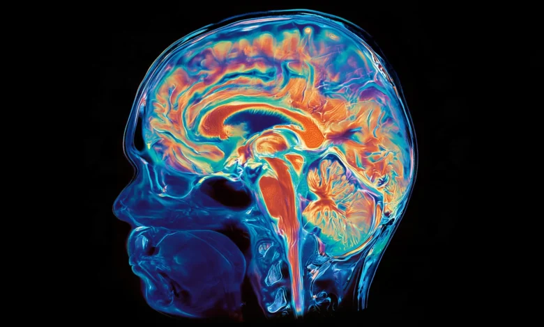
For decades, the prevailing doctrine in developmental biology has treated the human brain as a monolithic organ, a unified structure arising from a singular, ancestral cell population during the earliest stages of embryonic growth. This foundational model suggested that the forebrain, midbrain, and hindbrain were simply differentiated regions of one continuous developmental arc. However, groundbreaking new research published in the September 18 edition of Nature Neuroscience by a team at Stanford Medicine has fundamentally dismantled this assumption. The study reveals that the human brain is not a single, integrated creation, but rather a biological composite—a fusion of two distinct, ancient nervous systems that have evolved in parallel for over half a billion years.
The implications of this discovery are profound, offering a resolution to a long-standing conundrum in regenerative medicine: why scientists have historically struggled to cultivate specific brain stem neurons in laboratory settings. By identifying the separate progenitor lineages that give rise to the front and back of the brain, the research team, led by Associate Professor of Developmental Biology Kyle Loh, has unlocked a new method for generating hindbrain neurons, potentially opening the door to breakthrough therapies for debilitating neurodegenerative conditions such as Amyotrophic Lateral Sclerosis (ALS) and Spinal Muscular Atrophy (SMA).
A Reevaluation of Embryonic Gastrulation
The breakthrough originated from a meticulous examination of the earliest phases of embryonic development, specifically the period known as gastrulation. During this critical window, the body’s basic architecture is established as cells begin to commit to specific fates. Graduate students Carolyn Dundes and Rayyan Jokhai, the study’s co-first authors, spearheaded the investigation into how these precursor cells decide their ultimate destination.
Through the analysis of mouse embryos, the researchers identified two mutually exclusive populations of neural progenitor cells. The first population, characterized by the expression of the gene Otx2, is programmed to construct the forebrain and midbrain—the seats of high-level cognition, language, and abstract thought. The second population, marked by the expression of the gene Gbx2, is solely dedicated to forming the hindbrain, or brain stem, which governs autonomic life-support functions such as heart rate, respiration, and sleep cycles.
The team’s analysis of chromatin—the complex of DNA and proteins that determines gene accessibility—confirmed that these two populations are distinct from their inception. The chromatin configurations of the anterior neural ectoderm and the posterior neural ectoderm were fundamentally different, effectively locking these cell groups into separate evolutionary trajectories. This discovery explains why previous attempts to generate hindbrain neurons in petri dishes frequently failed; researchers were likely attempting to "reprogram" forebrain-destined cells into a hindbrain fate—a biological conversion that the study suggests is not possible under standard laboratory conditions.
Evolutionary Deep-Dive: 550 Million Years of Parallel Development
To understand the scope of this duality, the research team looked beyond human biology, conducting a comparative analysis spanning more than 550 million years of evolutionary history. The evidence of this "dual-origin" arrangement was found in a diverse array of species, including chickens, zebrafish, and even the simple acorn worm.
The most striking observation was found in jellyfish, organisms that diverged from the human lineage roughly 600 to 700 million years ago. These creatures possess two distinct nervous systems positioned at opposite ends of their bodies. This cross-species pattern suggests that the modern vertebrate brain is the result of evolution physically pushing two separate, pre-existing neural modules into close proximity. While the fusion has allowed for the highly efficient, integrated performance of the modern human brain, the "seams" of this merger remain visible in the distinct genetic lineages of our earliest development.
"Evolution took two existing neural systems and pushed them together spatially," said Dr. Loh. "Having the brain as one organ would arguably be more efficient, but we are still tethered to this primordial, two-piece construction method."
Addressing the Laboratory Bottleneck
The inability to grow functional hindbrain neurons has been a persistent "black box" for neuroscientists. Because the brain stem controls essential motor functions, the inability to replicate these cells has hampered the study of diseases like ALS, which affects the brain stem and spinal cord, and SMA, a leading genetic cause of infant mortality.
In both conditions, specific hindbrain neurons progressively degrade, leading to the loss of the ability to swallow—which increases the risk of aspiration pneumonia—and eventually the loss of respiratory function. Because brain stem tissue cannot be ethically or safely harvested from living patients for research, scientists have been forced to rely on animal models or limited post-mortem samples, neither of which fully capture the complex, age-related progression of human neurodegeneration.
By applying their discovery of the Gbx2-positive progenitor lineage, the Stanford team successfully guided human pluripotent stem cells into becoming functional hindbrain motor neurons. These lab-grown cells exhibited authentic behavioral markers, including the production of electrical action potentials and the synthesis of proteins critical for facial and swallowing muscles. This success represents a paradigm shift in stem cell research, as it underscores the necessity of focusing on early-stage developmental pathways rather than solely attempting to coax mature cell types into existence.
Clinical Implications and Future Research
The medical community has reacted with cautious optimism, noting that this research provides a viable roadmap for disease modeling. With the ability to grow patient-specific hindbrain neurons, researchers can now observe the cellular onset of ALS and SMA in real-time, screen potential drug candidates against these neurons, and test regenerative therapies with unprecedented precision.
Furthermore, the study touches upon the physiology of metabolism. The hindbrain is the regulatory center for hunger and satiety signals, areas of the brain that are increasingly targeted by the new generation of GLP-1 receptor agonist drugs used for weight management. Understanding the specific developmental origin of these circuits could lead to more targeted treatments for obesity and metabolic disorders.
"Now we have a model to better understand these devastating diseases, and work toward regenerative therapies for them," noted Rayyan Jokhai. "This is a very exciting new frontier in brain research."
The research team is already setting its sights on the next phase of inquiry: mapping the developmental origins of the spinal cord to see if it follows a similar parallel path. As scientists move forward, this work serves as a reminder that the human brain is not a static object of study, but a dynamic, layered system that bears the marks of deep evolutionary history. The "two-brain" model, while complex, provides a vital new lens through which to view human health, disease, and the long, segmented story of vertebrate evolution.
This ambitious study was supported by a wide coalition of organizations, including the National Institutes of Health, the National Science Foundation, and the California Institute for Regenerative Medicine, among others, reflecting the high stakes and broad interest in unlocking the mysteries of human neural development. As the field digests these findings, the focus will undoubtedly shift toward utilizing this newfound knowledge to alleviate the suffering caused by the very neural systems that have been, for so long, beyond our reach.