
Claude M. Steele Unveils "Churn": A Deep Dive into Identity Tension and the Power of Trust in Diverse Societies

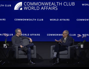
San Francisco, CA — In a compelling discussion at the Commonwealth Club World Affairs on March 11, 2026, Dr. Claude M. Steele, the eminent social psychologist and Professor Emeritus at Stanford University, introduced his new book, Churn: The Tension That Divides Us and How to Overcome It. This latest work serves as a profound sequel to his seminal 2010 publication, Whistling Vivaldi: How Stereotypes Affect Us and What We Can Do, which fundamentally reshaped the psychological understanding of prejudice and its effects. The event, featuring an on-stage interview with Jeremy Adam Smith, delved into the nuanced interplay of identity, anxiety, and trust in shaping human interactions across diverse settings.
The Enduring Legacy of Stereotype Threat
Before exploring the intricacies of Churn, Dr. Steele and Smith revisited the groundbreaking concept of stereotype threat, the cornerstone of Whistling Vivaldi. Steele, a former executive vice chancellor and provost at the University of California, Berkeley, explained that stereotype threat describes the phenomenon where individuals underperform in situations where they fear confirming a negative stereotype about their social group. This anxiety, often subtle yet potent, diverts cognitive resources from the task at hand, leading to suboptimal performance.
The research pioneered by Steele and his colleagues—including Steve Spencer and Josh Aronson—demonstrated how this psychological burden could account for significant disparities in academic test scores, workplace evaluations, and various other high-stakes environments. For instance, studies revealed that highly capable African American students, when confronted with challenging exams, might experience not only the normal frustrations of difficulty but also an additional layer of concern: Am I confirming a negative stereotype about my group? Will my performance be interpreted through that lens? This added pressure, the research showed, could tangibly undermine their performance. Similarly, women in STEM fields have been found to underperform on math tests when reminded of gender stereotypes about mathematical ability, despite having equal capabilities.
Whistling Vivaldi garnered widespread acclaim for its clear articulation of this pervasive dynamic, influencing fields from educational policy and curriculum design to organizational leadership and diversity training. Its insights provided a critical framework for understanding how systemic inequalities could perpetuate through psychological mechanisms, even in the absence of overt prejudice. The book’s impact was so significant that it became a foundational text for scholars and practitioners seeking to understand and mitigate barriers to equitable achievement.
From Individual Performance to Intergroup Tension: The Birth of "Churn"
While stereotype threat primarily focuses on the individual’s experience of underperformance, Steele recognized a broader implication: the same underlying tension often permeates interactions between people of different identities. This realization sparked the conceptual development of "churn," which he defines as a form of social anxiety tied to identity.
"Churn" describes the vigilant anxiety individuals experience when they worry about how their identity—be it race, age, gender, religion, or any other social category—will shape their interactions and how they will be judged and treated in diverse, important situations. It is a heightened awareness of potential negative stereotyping, leading individuals to become hyper-aware of their behavior, words, and interpretations.

Steele illustrated this with a vivid example: a seventh-grade parent-teacher conference involving African American parents and their son, and a white teacher. The African American parents, keenly aware of societal stereotypes regarding their racial identity, might enter the meeting with concerns: Will the teacher accurately perceive our child’s potential? Will ordinary mistakes be misconstrued as aggression or a lack of ability? They are in a state of churn, a vigilant anxiety about how their identities will influence the meeting’s outcome and their son’s school experience.
Crucially, Steele emphasized that churn is not limited to those from historically marginalized groups. The white teacher, deeply committed to fairness, might experience her own form of churn. Aware of stereotypes about her racial identity, she might worry that any constructive criticism could be misinterpreted as racism, leading to guarded communication. Both parties, despite potentially holding no personal prejudices, enter the interaction in a state of agitated concern about how their identities will affect their judgment and treatment. This distinguishes churn from traditional prejudice, as it arises from identity threat that all parties in a diverse setting can feel, regardless of their individual biases.
The Cognitive Burden of Churn: Multitasking and Flow Disruption
Jeremy Adam Smith aptly compared the experience of churn to "multitasking," a metaphor Steele readily endorsed. A person experiencing churn is not fully immersed in the primary activity or conversation; instead, they are simultaneously juggling extra mental tasks: monitoring their own behavior, anticipating others’ reactions, and constantly evaluating whether they are being perceived through a stereotypical lens. This cognitive load detracts from their ability to focus, engage authentically, and perform optimally.
In contrast, in homogenous groups—say, a group of older men—an individual might feel little anxiety about ageist stereotypes. However, the introduction of younger colleagues can trigger churn, prompting questions like: Do they think my ideas are outdated? Do they assume I’m technologically unsavvy? This loss of "security from outgroup stereotypes" is what generates the tension of churn. It prevents individuals from entering a state of "flow," where they are fully absorbed and performing at their best, instead keeping them in a state of vigilant self-monitoring. Churn, Steele clarified, isn’t inherently negative; it’s a natural coping mechanism in response to identity threat, signaling that the situation hasn’t yet fostered enough trust to feel safe.
Trust: The Antidote to Churn
The pivotal insight of Churn is that trust serves as the most powerful antidote to this identity-based anxiety. Steele detailed an illuminating experiment conducted with colleagues at Stanford University. White and Black students were asked to write essays about their favorite teacher, with the promise of potential publication. Two days later, they received feedback from a white evaluator.
When the feedback was delivered straightforwardly or preceded by generic praise, white students generally trusted it. Black students, however, exhibited significantly less trust. Their churn manifested as uncertainty: Did the criticism genuinely reflect the essay’s quality, or was it influenced by stereotypes about my group’s abilities?
The dynamic shifted dramatically when the evaluator prefaced the feedback with a specific message: "I’m applying high standards to these essays, and I believe you can meet those standards." This "wise feedback" transformed Black students’ trust, making them more trusting of the criticism than any other group. They were also far more likely to revise their essays based on this feedback. The message clearly signaled: "I am not judging you through negative stereotypes; I believe in your capability." This communication, Steele concluded, actively builds trust, thereby dissolving churn.
Building Trust at the Individual and Institutional Levels
Steele emphasized that building trust, at an individual level, often boils down to conveying genuine recognition of another person’s full humanity. He introduced the term "wise," borrowed from 1950s ethnographies of gay communities, referring to an outsider who understood and respected their humanity, refusing to reduce them to stereotypes.
For individuals, the simplest and most effective way to demonstrate this "wisdom" is through genuine curiosity: actively listening, asking open-ended questions, and taking a sincere interest in another’s experiences. This learning mindset, rather than a defensive or retreating posture, can profoundly transform interactions. While some might perceive this as "extra work," Steele countered that in high-stakes environments—classrooms, workplaces, teams—the effort invested in showing respect and interest is far less than the cost of dealing with the consequences of distrust and disengagement. Furthermore, he believes many people genuinely desire to bridge divides and would welcome concrete strategies to do so.
At an institutional level, building trust involves creating conditions where individuals feel psychologically safe and valued. Steele cited examples of university programs that successfully fostered environments where students genuinely trusted their institutions. In these settings, diversity transitioned from being a perceived "problem" to a "treasured feature" of the overall experience.
Navigating Power Dynamics and Societal Challenges
The discussion also tackled the complex issue of power dynamics. Steele acknowledged the inherent difficulty in expecting historically disempowered groups to initiate trust-building, particularly in a climate of heightened social and political division. He lamented the current era where political rhetoric often bypasses subtle "dog whistles" for openly prejudiced messaging, making it profoundly challenging for marginalized groups to trust that their full humanity is recognized or appreciated.
Despite this troubling reality, Steele urged against despair. His book, he clarified, isn’t about solving grand political issues directly but about empowering individuals and institutions within the diverse settings of their everyday lives. "We still have to get up every day and go to work," he stated, highlighting the practical need for strategies that enable people to feel more comfortable and engaged across identity divides, thereby unlocking the "riches that our differences can offer us."
Steele candidly admitted to experiencing churn himself, particularly as an older individual working in environments dominated by younger people. He shared moments where he’d wonder if he was being stereotyped as technologically inept. This personal anecdote underscored the universality of churn, affirming that it’s a shared human experience that transcends specific group affiliations.
The American Experiment and a Path Forward

Jeremy Adam Smith noted the "neurotic" aspect of the American experiment, simultaneously desiring a diverse society and fearing its implications, grappling with the tension between the comfort of sameness and the vitality of difference. Steele concurred, emphasizing that this inherent challenge is precisely why focusing on tangible, local contexts—classrooms, workplaces, hiring systems—is crucial. Abstract discussions about "larger society" can feel utopian, but concrete strategies applied in specific settings can yield real progress.
He drew a historical parallel to the 1960s, when the U.S. dismantled segregation laws, making a legal commitment to a multiracial democracy. While that was a monumental achievement, the ongoing challenge is to realize "equality of opportunity in everyday life," which primarily unfolds through relationships and the "small things people do that make it easier to trust each other and work together."
Steele expressed optimism that building trust might be a more manageable and effective approach than directly trying to eliminate deep-seated prejudices. "Changing someone’s beliefs is incredibly hard," he noted, citing his decades of experience as a social psychologist. "But trust is different. Many of us have done that in our lives. We have fairly good intuitions about how to do it and what’s required." He posited that as trust builds, attitudes and beliefs can naturally begin to shift, and prejudices may loosen their grip in the presence of genuine human connection.
A Call for New Research
Concluding the interview, Steele articulated his hope for future research spurred by Churn. Just as Whistling Vivaldi ignited years of inquiry into stereotype threat, he desires to see extensive research dedicated to testing whether a deliberate focus on building trust is an effective means of reducing prejudice. He believes this trust-building approach has been an "under-appreciated factor" in bridging identity differences.
Steele recounted instances where educators, for example, expressed frustration that students "don’t listen" despite clear explanations. He argued that if students are wary of trusting the educator, information alone is insufficient. Instead, they first need "some evidence or signal that their full humanity is appreciated—that they’re not being reduced to those stereotypes that they know exist, and that they know you know." Once this foundation of trust is established, the path to learning and productive interaction becomes significantly smoother.
Dr. Claude M. Steele’s Churn is poised to become another essential contribution to social psychology, offering not only a profound diagnostic framework for understanding the tensions in diverse interactions but also a practical, hopeful roadmap for building trust and fostering more inclusive, equitable, and cohesive societies. His work encourages individuals and institutions alike to seriously explore the "road of trust-building" in the diverse settings that constitute our daily lives.


